

 PDF
PDF ePub
ePub Citation
 Print
Print


Abstract
Purpose:
To review the clinical manifestations, indications and the management outcomes of adult patients with ureteroceles.
Materials and Methods:
Between 1995 and 2006, 20 adult patients (9 females, 3 males) with ureteroceles were investigated for their clinical symptoms, type of ureterocele and renal function. The outcomes of surgical or conservative management, according to the patients’ symptoms were also individually analyzed. The median follow-up was 38 months (12-50 months).
Results:
The ages at diagnosis of the ureteroceles ranged from 19 to 70 years (mean 37.9 years). The ureterocele-related symptoms were flank pain (3), hematuria (1) and lower urinary tract symptoms (4). Two cases were incidentally detected with ultrasound (1) or computed tomography (1), and another 2 patients presented with non-specific flank pain or a hematuria. Eight patients exhibited an intravesical single system and 4 were associated with upper pole of a duplex system. Only one patient had an ectopic ureterocele, in which the orifice was located in the mid-urethra. The ureterocele-related symptoms were managed using a transurethral incision (5) or resection (1) of the ureterocele, with ure- teroscopic stone retrieval (2). The symptoms were resolved after surgery, and there were no recurrence of symptoms or any deterioration of the renal function during follow-up.
Conclusions:
To diagnose an ureterocele in adult patients requires a high index of suspicion, as not all patients present with the typical clinical manifestations associated in children. Our results suggested that ure- terocele-related symptoms are the main indication for surgery in adult patients. While methods with lower morbidity may be a useful, expectant treatment, they may also be an appropriate option for the management of incidentally detected ureteroceles. ι
Go to : 
REFERENCES
1.Coplen DE., Duckett JW. The modem approach to ureteroceles. J Urol. 1995. 153:166–71.
2.Synder HM. Anomalies of the ureter. Gillenwater JY, Grayhack JT, Howard SS, Duckett JW, editors. editors.Adult and pediatric urology. 2nd ed.Chicago: Mosby-Year Book;1991. p. 1831–62.
3.Song C., Park YS., Kim KS. Clinical outcome of various surgical methods for the ureteroceles in children. Korean J Urol. 2005. 46:823–8.
4.Van Savage JG., Mesrobian HG. The impact of prenatal sonography on the morbidity and outcome of patients with renal duplication anomalies. J Urol. 1995. 153:768–70.

5.Hulbert WC., Rabinowitz R. Prenatal diagnosis of duplex system hydronephrosis: effect on renal salvage. Urology. 1998. 51:23–6.
6.Glassberg KI., Braren V., Duckett JW., Jacobs EC., King LR., Lebowitz RL, et al. Suggested terminology for duplex system, ectopic ureters and ureteroceles. J Urol. 1984. 132:1153–4.
7.Sen S., Beasley SW., Ahmed S., Smith ED. Renal function and vesicoureteric reflux in children with ureteroceles. Pediatr Surg Int. 1992. 7:192–4.
8.Amitai M., Hertz M., Jonas P., Apter S., Heyman Z. Ectopic ureterocele in adults with a comparison of the anomaly in children. Urol Radiol. 1992. 13:181–6.
9.Feldman S., Lome L. Surgical management of ectopic ureterocele. Urology. 1981. 17:252–6.
10.Albers P., Foster RS., Bihrle R., Adams MC., Keating MA. Ectopic ureters and ureteroceles in adults. Urology. 1995. 45:870–4.
11.Griffin J., Jennings C., MacErlean D. Ultrasonic evaluation of simple and ectopic ureterocele. Clin Radiol. 1983. 34:55–7.
12.Schwartz ML., Kenney PJ., Bueschen AJ. Computed tomographic diagnosis of ectopic ureter with seminal vesicle cyst. Urology. 1988. 31:55–6.
13.Schlussel RN., Retik AB. Anomalies of the ureter. In:. Walsh PC, Retik AB, Vaughan ED, Wein AJ, editors. editors.Campbell' Surology. 7th ed.Philadelphia: Saunders;1998. p. 1831–2.
14.Han MY., Gibbons MD., Belman AB., Pohl HG., Majd M., Rushton HG. Indications for nonoperative management of ureteroceles. J Urol. 2005. 174:1652–6.
15.Shankar KR., Vishwanath N., Rickwood AM. Outcome of patients with prenatally detected duplex system ureterocele; natural history of those managed expectantly. J Urol. 2001. 165:1226–8.
Go to :
 | Fig. 1.Computed tomography showing a right atrophied kidney (arrow) associated with an ipsilateral ureterocele. |
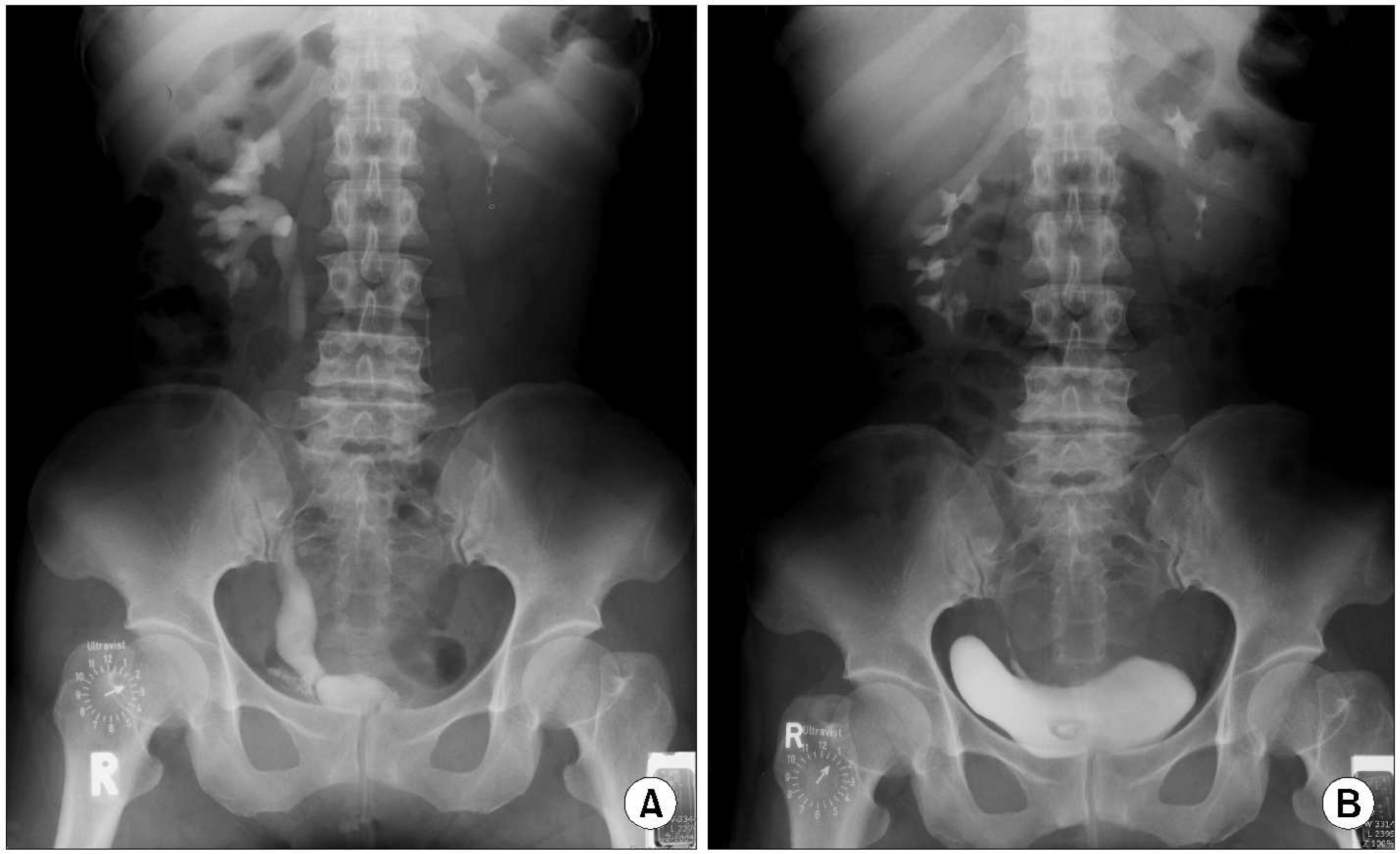 | Fig. 2.Intravenous pyelography (IVP) of a 40-year-old female, who presented with urinary interruption of 3 years duration. (A) Preoperative IVP showing an intravesical ureterocele of a single system. (Β) Decreased size of the ure- terocele after a transurethral incision of the ureterocele. |
Table 1.
Patients’ characteristics
IVP: intravenous pyelography, TUI: transurethral incision of ureterocele, URS: ureteroscopic retrieval of stone, UVJ: ureterovesical junction, TUR: transurethral resection of ureterocele, Recurrent UTI: recurrent urinary tract infection, ∗Duplex: duplex type ureterocele with ectopic orifice opening to mid-urethra, CT: computed tomography
 XML Download