

 PDF
PDF ePub
ePub Citation
 Print
Print


Abstract
Purpose
The accurate analysis of urinary stone components is fundamental for studying of the etiology of stone formation and it is essential for the treatment of urinary stone and its prevention. We compared the analysis of urinary stone components during the last two decades.
Materials and Methods
Stone analysis was performed by Louis C. Herring and Company. The urinary stones were aobtained from January, 1986 to December, 2005. We compared the stone components of the first decade (Group A, 301 cases) with that of the second decade (Group B, 158 cases).
Results
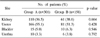
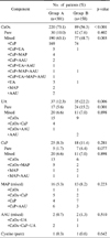
The mean age was 47.2±14.5 in Group A and 46.0±10.2 in Group B (p=0.658). The ratio of males to females was 2.04:1 in Group A and 1.98:1 in Group B (p=0.888). Ureteral stones were the most common stones in both groups. Among all the components analyzed in Group A, calcium oxalate made up 73.1% of the total. Other components found in the stones were uric acid 12.3%, calcium phosphate 8.3%, magnesium ammonium 5.3%, ammonium acid urate 0.7% and cystine 0.3%. In Group B, calcium oxalate was only 56.3% of the total and uric acid was 22.2%, calcium phosphate was 11.4%, magnesium ammonium phosphate was 8.2%, ammonium acid urate was 1.3% and cystine was 0.6%. On comparison of the stones of the two groups, the incidence of calcium oxalate was decreased in Group A (73.1% vs 56.3%, p<0.001). However, the incidence of uric acid in Group B was increased (12.3% vs 22.2%, respectively, p=0.006). There were no notable statistical increases in the frequency of the other components (p>0.05).
Figures and Tables
References
1. Leusmann DB, Blaschke R, Schmandt W. Results of 5,035 stone analyses: a contribution to epidemiology of urinary stone disease. Scand J Urol Nephrol. 1990. 24:205–210.
2. Johnson CM, Wilson DM, O'Fallon WM, Malek RS, Kurland LT. Renal stone epidemiology: a 25-year study in Rochester, Minnesota. Kidney Int. 1979. 16:624–631.
3. Borghi L, Schianchi T, Meschi T, Guerra A, Allegri F, Maggiore U, et al. Comparison of two diets for the prevention of recurrent stones in idiopathic hypercalciuria. N Engl J Med. 2002. 346:77–84.
4. Uribarri J, Oh MS, Carroll HJ. The first kidney stone. Ann Intern Med. 1989. 15:1006–1009.
5. Lee SJ, Kim DK, Rho SK, Huh JS, Lee HL, Lee CH, et al. Clinical observations in 4,468 cases of patients with urinary stones. Korean J Urol. 1996. 37:877–887.
6. Hiatt RA, Dales LG, Friedman GD, Hunkeler EM. Frequency of urolithiasis in a prepaid medical care program. Am J Epidemiol. 1982. 115:255–265.
7. Stamatelou KK, Francis ME, Jones CA, Nyberq LM, Curhan GC. Time trends in reported prevalence of kidney stones in the United States: 1976-1994. Kidney Int. 2003. 63:1817–1823.
8. Yu T, Gutman AB. Uric acid nephrolithiasis in gout. Predisposing factors. Ann Intern Med. 1967. 67:1133–1148.
9. Asper R. Epidemiology and socioeconomic aspects of urolithiasis. Urol Res. 1984. 12:1–5.
10. Balaji KC, Menon M. Mechanism of stone formation. Urol Clin North Am. 1997. 24:1–11.
11. Menon M, Mahle CJ. Oxalate metabolism and renal calculi. J Urol. 1982. 127:148–151.
12. Finlayson B. Symposium on renal lithiasis. Renal lithiasis in review. Urol Clin North Am. 1974. 1:181–212.
13. Byeon SS, Kim HH, Kim SW. Analysis of the urinary stone components using chemical analysis method. Korean J Urol. 1996. 37:179–186.
14. Hossain RZ, Ogawa Y, Hokama S, Morozumi M, Hatano T. Urolithiasis in Okinawa, Japan: a relatively high prevalence of uric acid stones. Int J Urol. 2003. 10:411–415.
15. Koide T, Itatani H, Yoshioka T, Ito H, Namiki M, Nakano E, et al. Clinical manifestations of calcium oxalate monohydrate and dihydrate urolithiasis. J Urol. 1982. 127:1067–1069.
16. Prien EL, Frondel C. Studies in urolithiasis: I, The composition of urinary calculi. J Urol. 1947. 57:949–994.
17. Prien EL. Composition and structure of urinary stone. Am J Med. 1968. 45:654–672.
18. Kim CS, Kim CG. Analysis of urinary calculi by X-ray diffraction method. Korean J Urol. 1987. 28:233–245.
19. Pak CY, Skurla C, Harvey J. Graphic display of urinary risk factors for renal stone formation. J Urol. 1985. 134:867–870.
20. Asplin JR. Uric acid stones. Semin Nephrol. 1996. 16:412–424.
21. Matzkies F, Berg G. The uricosuric action of amino acids in man. Adv Exp Med Biol. 1977. 76:36–40.
22. Pak CH, Oleneva VA, Agadzhanov SA. Dietetic aspects of preventing urolithiasis in patients with gout and uric acid diathesis. Vopr Pitan. 1985. 1:21–24.
23. Herring LC. Observations on the analysis of ten thousand urinary calculi. J Urol. 1962. 88:545–562.
24. Klohn M, Bolle JF, Reverdin NP, Susini A, Baud CA, Graber P. Ammonium urate urinary stones. Urol Res. 1986. 14:315–318.
 XML Download