

 PDF
PDF ePub
ePub Citation
 Print
Print


Abstract
A 69-year-old man was admitted to the hospital because of flu-like symptoms and fatigue for 2 weeks. Computed tomography revealed ground glass opacity and consolidation in both the lungs as well as pleural effusion. The patient was diagnosed with pneumonia and was hospitalized. At the time of hospitalization, he complained of shortness of breath and coughed-up blood-tinged sputum. Two days after admission, he died suddenly. An autopsy was performed; cardiomegaly was noted, and further examination revealed that the aortic valve had been destroyed by multiple, irregular vegetations. Herein, we report an autopsy case of infective endocarditis with a review of the relevant literatures.
Go to : 
REFERENCES
1. Karchner AW. Infective endocarditis. Kasper DL, Braunwald E, Fauci AS, editors. ed.Harrison’ s principles of internal medicine. 16th ed.New York: McGraw-Hill;2005. p. 731–40.
2. Mylonakis E, Calderwood SB. Infective endocarditis in adults. N Engl J Med. 2001; 345:1318–30.

3. Na JY, Park JP, Park HJ, et al. The statistical analysis on legal autopsy performed in Korea during 2012 Year. Korean J Leg Med. 2013; 37:198–207.
4. Byramji A, Gilbert JD, Byard RW. Sudden death as a complication of bacterial endocarditis. Am J Forensic Med Pathol. 2011; 32:140–2.
5. Zeller L, Flusser D, Shaco-Levy R, et al. A rare complication of infective endocarditis: left main coronary artery embolization resulting in sudden death. J Heart Valve Dis. 2010; 19:225–7.
6. Saad R, Yamada AT, Pereira da Rosa FH, et al. Comparison between clinical and autopsy diagnoses in a cardiology hospital. Heart. 2007; 93:1414–9.
7. Ferna ′ndez Guerrero ML, A′lvarez B, Manzarbeitia F, et al. Infective endocarditis at autopsy: a review of pathologic manifestations and clinical correlates. Medicine (Baltimore). 2012; 91:152–64.
8. Cohle SD, Graham MA, Sperry KL, et al. Unexpected death as a result of infective endocarditis. J Forensic Sci. 1989; 34:1374–86.
Go to :
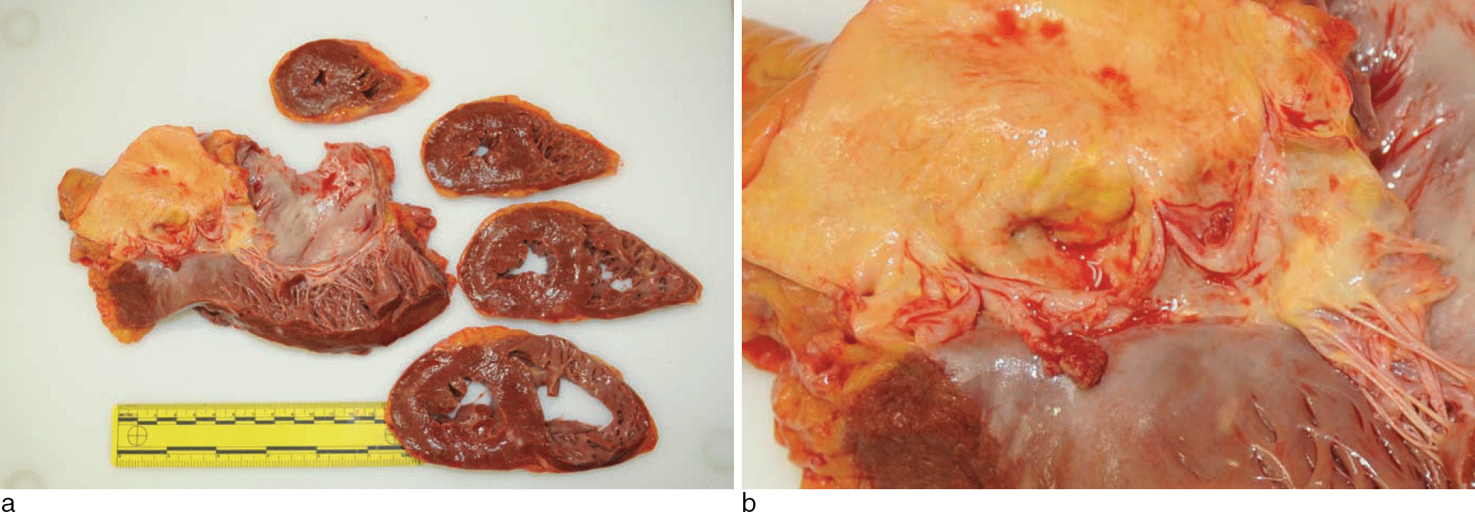 | Fig. 1.Destroyed aortic cusps and vegetations, measuring up to 1.2 × 0.7 ㎝ are noted. Cross sections of the myocardium show multiple patch-like fibrotic lesions. |
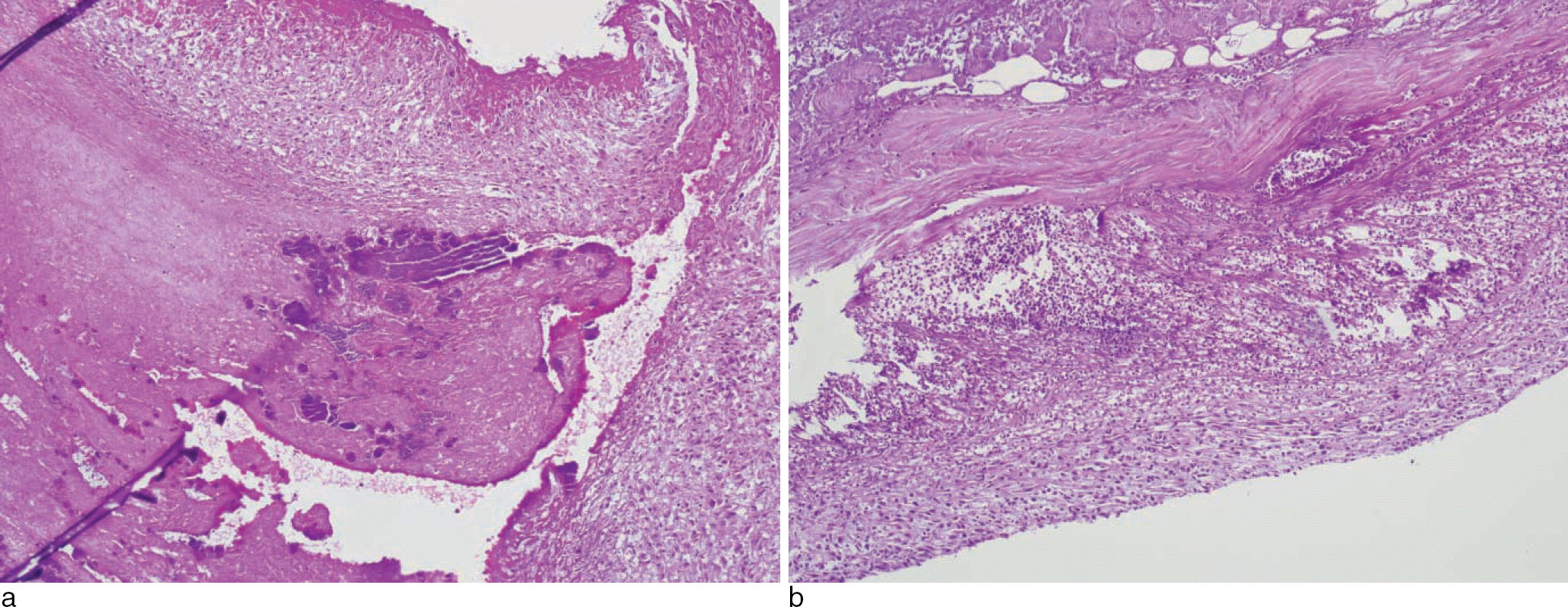 | Fig. 2.Microscopically, the aortic valve demonstrated friable vegetations comprising fibrin and platelets mixed with inflammatory cells and bacteria (H&E, × 100). |
Table 1.
Modified Duke Criteria for the Diagnosis of Infective Endocarditis2)
Table 2.
Causes of Unexpected Death in Patient with of Infective Endocarditis4)
 XML Download