Abstract
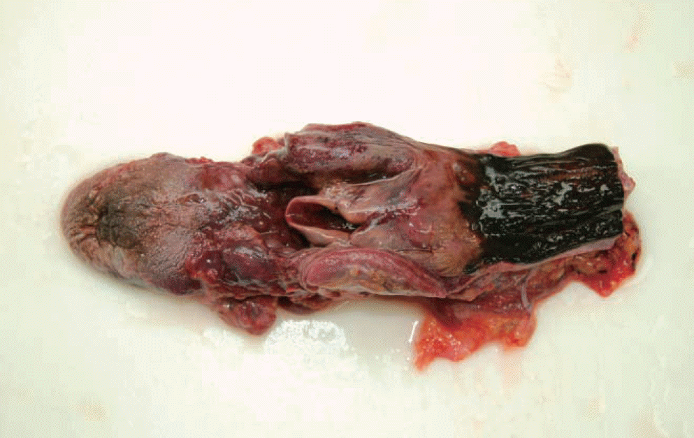
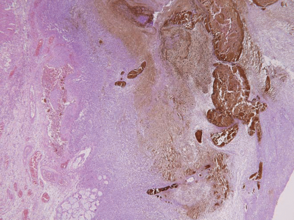
Acute necrotizing esophagitis (AEN), also called “black esophagus,” is a rare disorder with an unknown pathogenesis. Endoscopic findings generally show black pigmentation throughout the esophagus. This case also offered rare views of the gross anatomy of this disorder. Histological examination revealed that the mucosal and submucosal layers of the esophagus were involved in the severe necrotizing inflammation. The chief manifestation of this disease is hematemesis from hemorrhage of the upper gastrointestinal tract with a typically multifactorial etiology. AEN is also characterized by a clear boundary at the gastroesophageal junction where the necrosis stops. In this study, we report an autopsy case of a 61-year-old man with necrotizing inflammation throughout the esophagus and esophageal necrosis from the laryngopharynx to the gastroesophageal junction. The patient was a disabled person with a history of alcohol abuse who was also diagnosed with mild coronary arteriosclerosis and fatty liver on the basis of the underlying diseases. In this case, the main etiology for poor perfusion from the distal esophageal area was likely underlying illness, history of alcoholism, and malnutrition.
Go to : 
REFERENCES
2. Day A, Sayegh M. Acute oesophageal necrosis: a case report and review of the literature. Int J Surg. 2010; 8:6–14.

3. Choi EJ, Lee SH, Lee SH. A case of acute necrotizing esophagitis. Korean J Gastroenterol. 2010; 56:373–6.

4. Endo T, Sakamoto J, Sato K, et al. Acute esophageal necrosis caused by alcohol abuse. World J Gastroenterol. 2005; 11:5568–70.

5. Burtally A, Gregoire P. Acute esophageal necrosis and low-flow state. Can J Gastroenterol. 2007; 21:245–7.

6. Ramos R, Mascarenhas J, Duarte P, et al. Acute esophageal necrosis: a retrospective case series. Rev Esp Enferm Dig. 2008; 100:583–5.
7. Gurvits GE, Shapsis A, Lau N, et al. Acute esophageal necrosis: a rare syndrome. J Gastroenterol. 2007; 42:29–38.

8. Hwang J, Weigel TL. Acute esophageal necrosis: “black esophagus”. JSLS. 2007; 11:165–7.
9. Julia′n Go ′mez L, Barrio J, Atienza R, et al. Acute esophageal necrosis. An underdiagnosed disease. Rev Esp Enferm Dig. 2008; 100:701–5.
10. Notario A, Pallavicini EB, Marciano` S, et al. Blood and liver fat values in rats kept on a hyperlipid, hypoprotein, steatogenous diet, with or without choline. Arch Sci Med (Torino). 1973; 130:169–87.
Go to : 
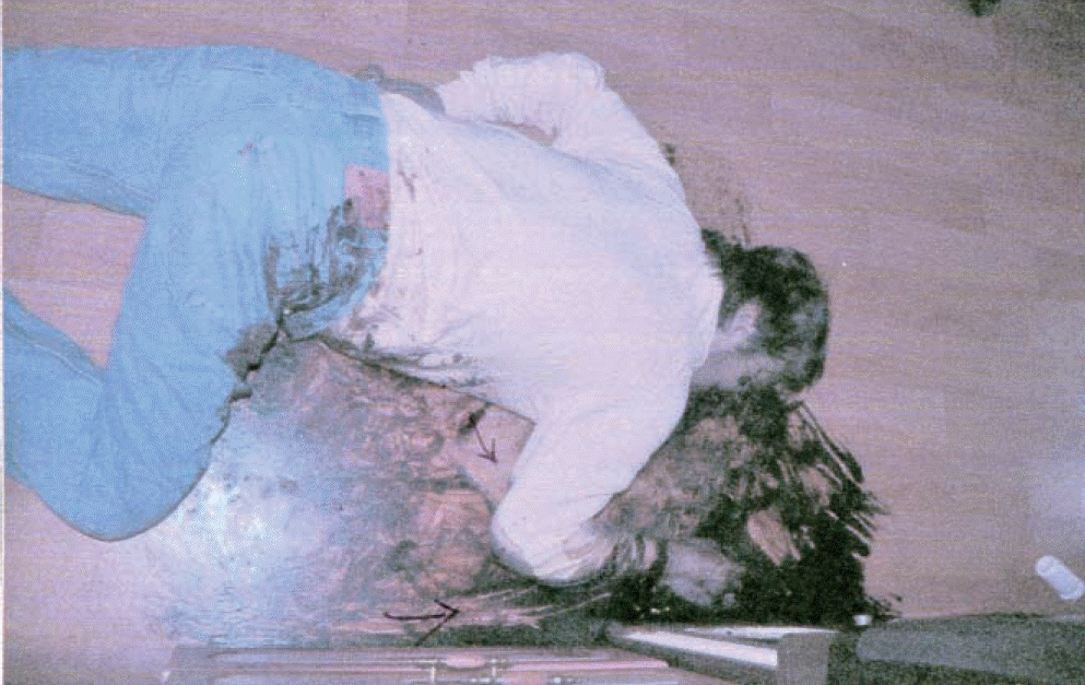
 | Fig. 1.The scene investigation showing that the floor around the deceased was soaked with coagulated blood and various writhing traces were made on it. |
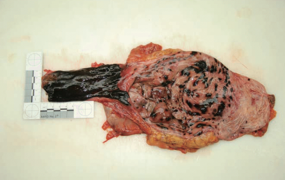
 | Fig. 2.Gross view of necrotizing inflammation of lower esophagus. The black esophagus was terminated at the esophagogastric junction, abruptly. The suspected erosive lesion on decomposed gastric mucosa is also observed. |


 PDF
PDF ePub
ePub Citation
 Print
Print


 XML Download