Abstract
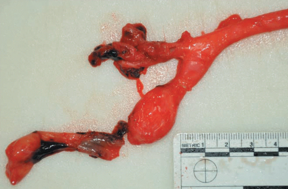
Aneurysm of the internal carotid artery is a rare disease and is known to be associated with congenital arterial anomalies such as neurofibromatosis type Ⅰ (NF-Ⅰ). NF-Ⅰ is an autosomal dominant neurocutaneous disorder characterized by a variety of manifestations that involve the central and peripheral nervous systems, skin, vascular system, and skeleton. In particular, the involvement of vascular abnormalities in NF-Ⅰ is well known. Any vessel may be affected by this condition, although the renal artery is most frequently involved. The vascular abnormality can be occlusive or an aneurysmal degenerative change. Therefore, symptomatic presentations might assume an indolent pathophysiologic course such as hypertension, or manifest as a catastrophic event such as arterial rupture that could result in sudden death. We report a rare autopsy case of an aneurysmal rupture of the internal carotid artery in a woman with suspected NF-Ⅰ, who collapsed in her home.
REFERENCES
1. Lee MJ, Stephenson DA. Recent developments in neurofibromatosis type 1. Curr Opin Neurol. 2007; 20:135–41.

2. Byard RW. Forensic considerations in cases of neurofibromatosis-an overview. J Forensic Sci. 2007; 52:1164–70.

3. Onkendi E, Moghaddam MB, Oderich GS. Internal carotid artery aneurysms in a patient with neurofibromatosis type 1. Vasc Endovascular Surg. 2010; 44:511–4.

4. McCollum CH, Wheeler WG, Noon GP, et al. Aneurysms of the extracranial carotid artery. Twenty-one years’ experience. Am J Surg. 1979; 137:196–200.
5. Reubi F. Neurofibromatose et lesions vasculaires. Schweiz Med Wochenschr. 1945; 75:463–5.
6. Hamilton SJ, Friedman JM. Insights into the pathogenesis of neurofibromatosis 1 vasculophathy. Clin Genet. 2000; 58:341–4.
7. Friedman JM, Arbiser J, Epstein JA, et al. Cardiovascular disease in neurofibromatosis 1: report of the NF1 cardiovascular task force. Genet Med. 2002; 4:150–11.

8. Oderich GS, Sullivan TM, Bower TC, et al. Vascular abnormalities in patients with neurofibromatosis syndrome type Ⅰ: clinical spectrum, management, and results. J Vasc Surg. 2007; 46:475–84.
Fig. 1.
Postmortem examination shows no evidence of external injury, but swelling and subcutaneous hemorrhage in the right neck and shoulder region are noted.
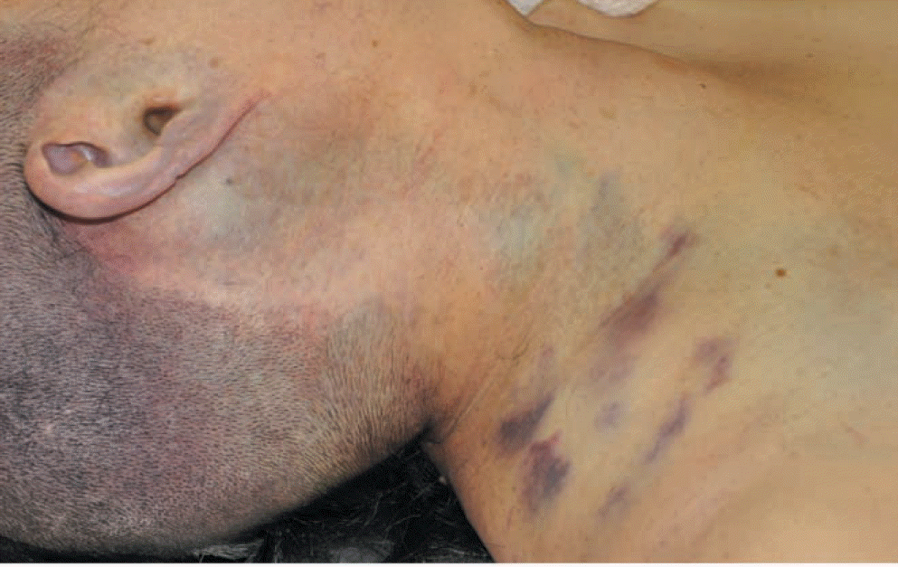
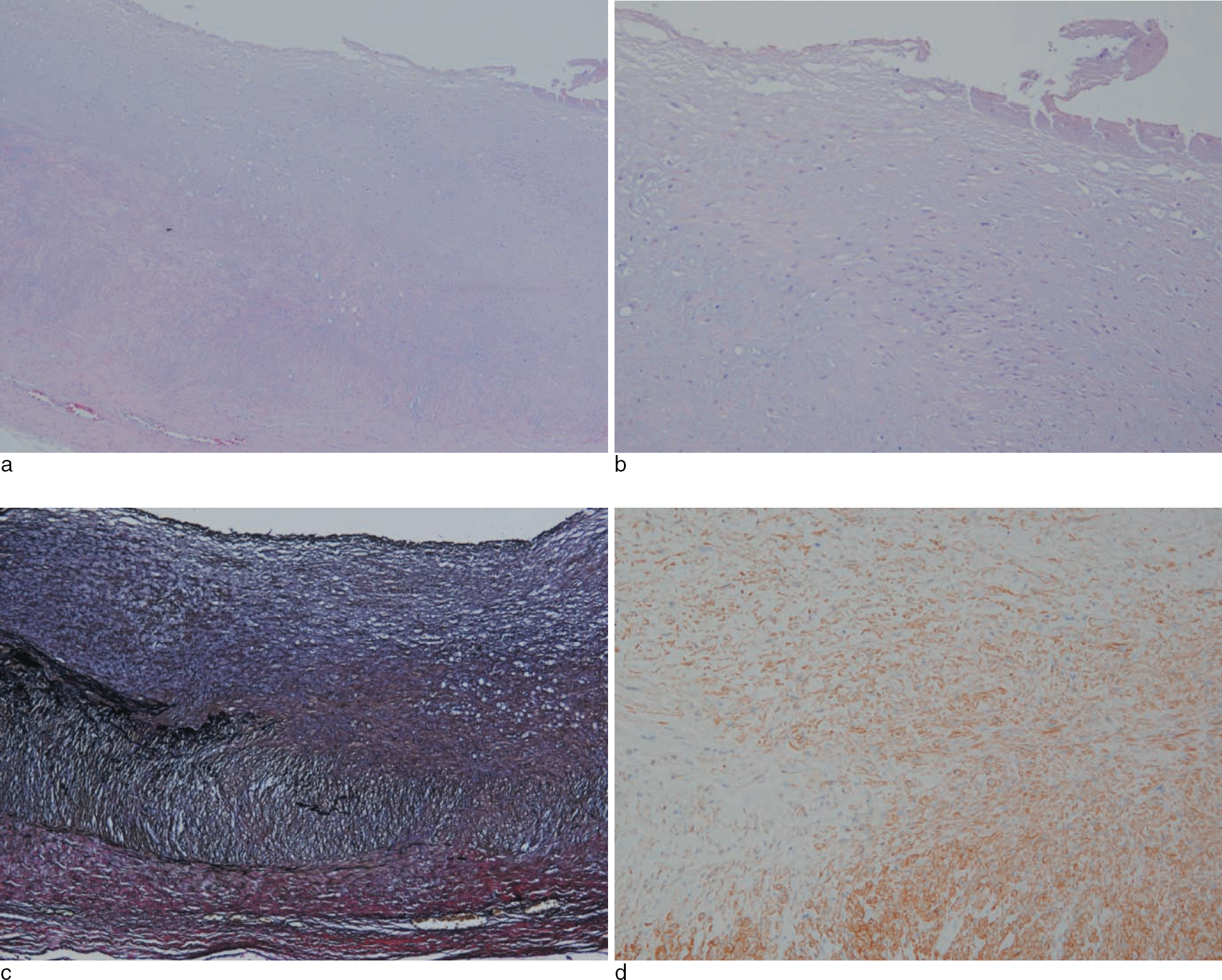
Fig. 5.
Disrupted elastic lamina and thickened tunica intima are noted accompanying spindle cell proliferation (Fig. 5a, H&E, x40; 5b, H&E, x100; 5c, elastic stain, x40). Proliferating spindle cells which are positive on the smooth muscle actin (SMA) are presented in the tunica intima of the right internal carotid artery (Fig. 5d, SMA, x100).


 PDF
PDF ePub
ePub Citation
 Print
Print


 XML Download