

 PDF
PDF ePub
ePub Citation
 Print
Print


Abstract
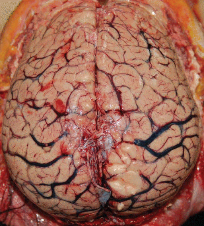
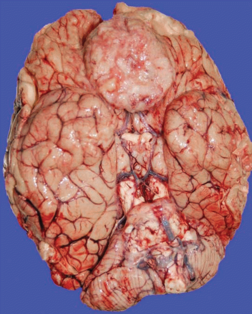
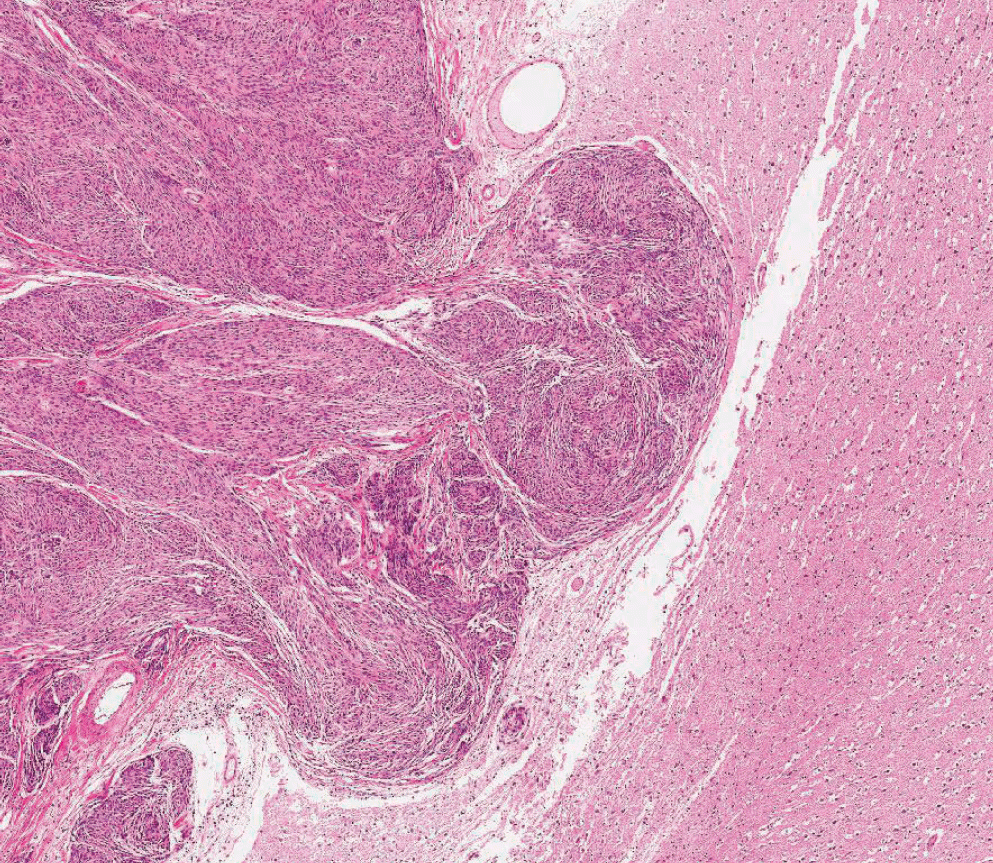
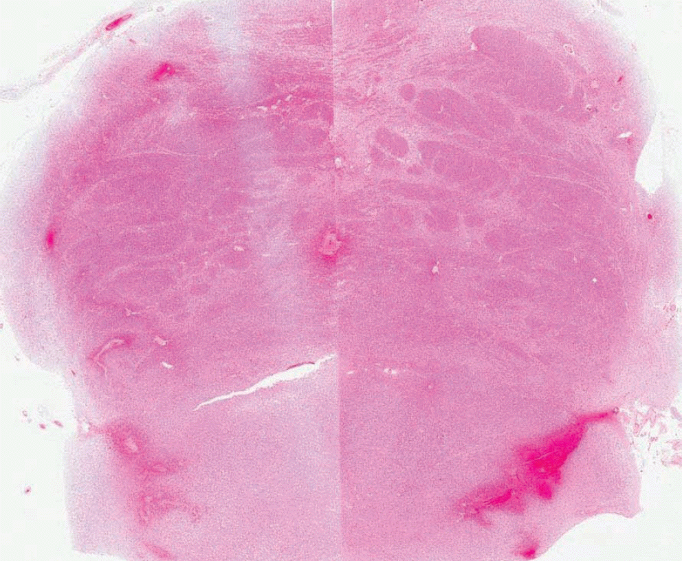
Meningiomas, one of the most common neoplasms of the central nervous system, may be encountered incidentally during autopsy. Most of these tumors, however, are benign and hence, are not considered as the chief cause of death. Further, sudden unexpected death caused by meningioma is very unusual. Moreover, the diagnosis of an incidental meningioma as the cause of sudden death may sometimes be difficult. In the present report, we describe an autopsy case of a sudden, unexpected death due to a large olfactory groove meningioma accompanied by severe cerebral edema and tonsillar herniation.
REFERENCES
1. Perry A, Louis DN, Scheithauer BW, et al. Menigiomas. Louis DN, Ohgaki H, Wiestler OD, editors. WHO classification of tumors of the central nervous system. Lyon: International Agency for Research on Cancer;2007. p. 164–72.
2. Nakamura M, Roser F, Michel J, et al. The natural history of incidental meningiomas. Neurosurgery. 2003; 53:62–70.

3. Nakano T, Asano K, Miura H, et al. Meningioma with brain edema: radiologic characteristics on MRI and review of the literature. Clin Imaging. 2002; 26:243–9.
4. Rausing A, Ybo W, Stenflo J. Intracranial meningioma– a population study of ten years. Acta Neurol Scand. 1970; 46:102–10.
5. Nakasu S, Hirano A, Shimura T, et al. Incidental meningiomas in autopsy study. Surg Neurol. 1987; 27:319–22.
6. Ohaegbulam SC. Sudden death from an asymptomatic sphenoid ridge meningioma. J Neurol. 1977; 215:291–4.
7. Lee BW, Seo JS. Meningioma presenting as cerebral infarct: case report. Korean J Leg Med. 2001; 25:53–7.
8. Huh GY, Kim KH, Ahn YW, et al. Sudden death due to undiagnosed intracranial meningioma: a case report. Korean J Leg Med. 2008; 32:150–2.
9. Helle TL, Conley FK. Haemorrhage associated with meningioma: a case report and review of the literature. J Neurol Neurosurg Psychiatry. 1980; 43:725–9.
10. Leestma JE, Kirkpatrick JB. Forensic neuropathology. 1st ed.New York: Raven;1988. p. 157–83.
 XML Download