

 PDF
PDF ePub
ePub Citation
 Print
Print


Abstract
Thyrotoxicosis (thyroid crisis) is a known cause of sudden death; however, only a few cases of death resulting from thyrotoxicosis have been reported. Histopathologic examination and postmortem thyroid function tests may be helpful in postmortem diagnosis, but their usefulness seems to be limited. We report three autopsy cases associated with thyrotoxicosis.
Go to : 
REFERENCES
1. James JL, Weetman AP. Disorders of the thyroid gland. Longo DL, Fauci AS, Kasper DL, editors. Harrison’ s principles of internal medicine. 18th ed.New York: McGraw-Hill;2011. p. 2911–39.
2. Franklyn JA, Boelaert K. Thyrotoxicosis. Lancet. 2012; 379:1155–66.

3. Salvatore D, Davies TF, Schilumberger M, et al. Thyrotoxicosis. Melmed S, Polonsky KS, Larsen PR, editors. Williams textbook of endocrinology. 11th ed.Philadelphia: Elsevier Sauders;2011. p. 362–405.
4. Nakatani Y, Monden T, Sato M, et al. Severe hypoglycemia accompanied with thyroid crisis. Case Rep Endocrinol. 2012 Nov 4. [Epub].http://dx.doi.org/10.1155/2012/168565.
5. Edston E, Druid H, Holmgren P, et al. Postmortem measurements of thyroid hormones in blood and vitreous humor combined with histology. Am J Forensic Med Pathol. 2001; 22:78–83.
6. Osman F, Franklyn JA, Holder RL, et al. Cardiovascular manifestations of hyperthyroidism before and after antithyroid therapy: a matched case-control study. J Am Coll Cardiol. 2007; 49:71–81.
7. Eberhart CG, Wiestler OD, Eng C. Cowden disease and dysplastic gangliocytoma of the cerebellum/Lhermitte-Duclos disease. Louis DN, Ohgaki H, Wiestler OD, editors. WHO Classification of Tumours of the Central Nervous System. Lyon: IARC Press;2007. p. 226–8.
8. Kwon TJ, Kim TS, Lee HY, et al. Lhermitte-Duclos disease in a sudden death: an autopsy case. Korean J Pathol. 1994; 28:73–8.
Go to :
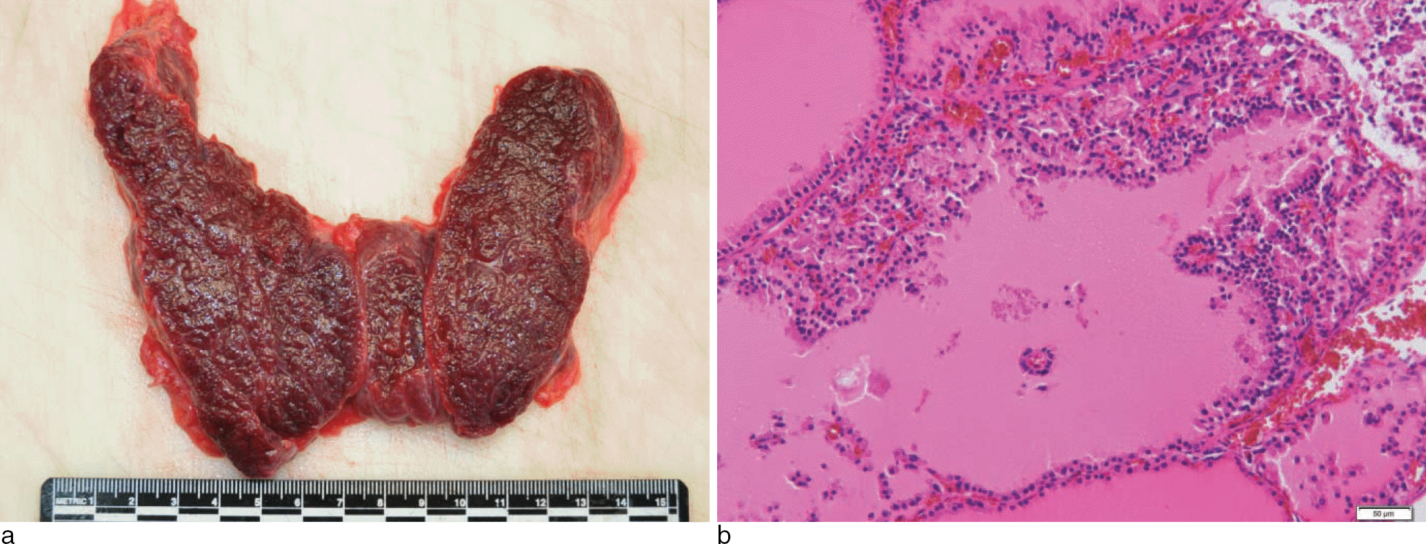 | Fig. 1.(Case 1) The thyroid gland shows relatively symmetric enlargement with soft and meaty parenchyma (a). There are follicles of varying size, lined by tall columnar cells with decrease in the amount of colloid and formation of pseudopapillary structures (b: H & E, × 200). |
 XML Download