Abstract
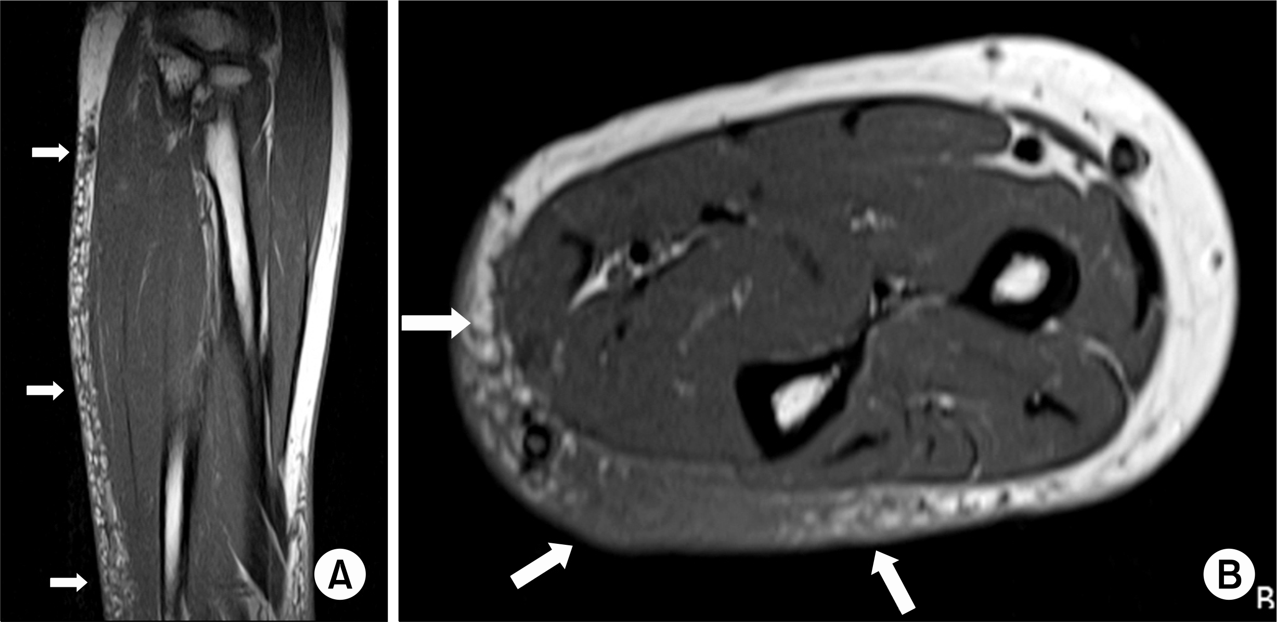
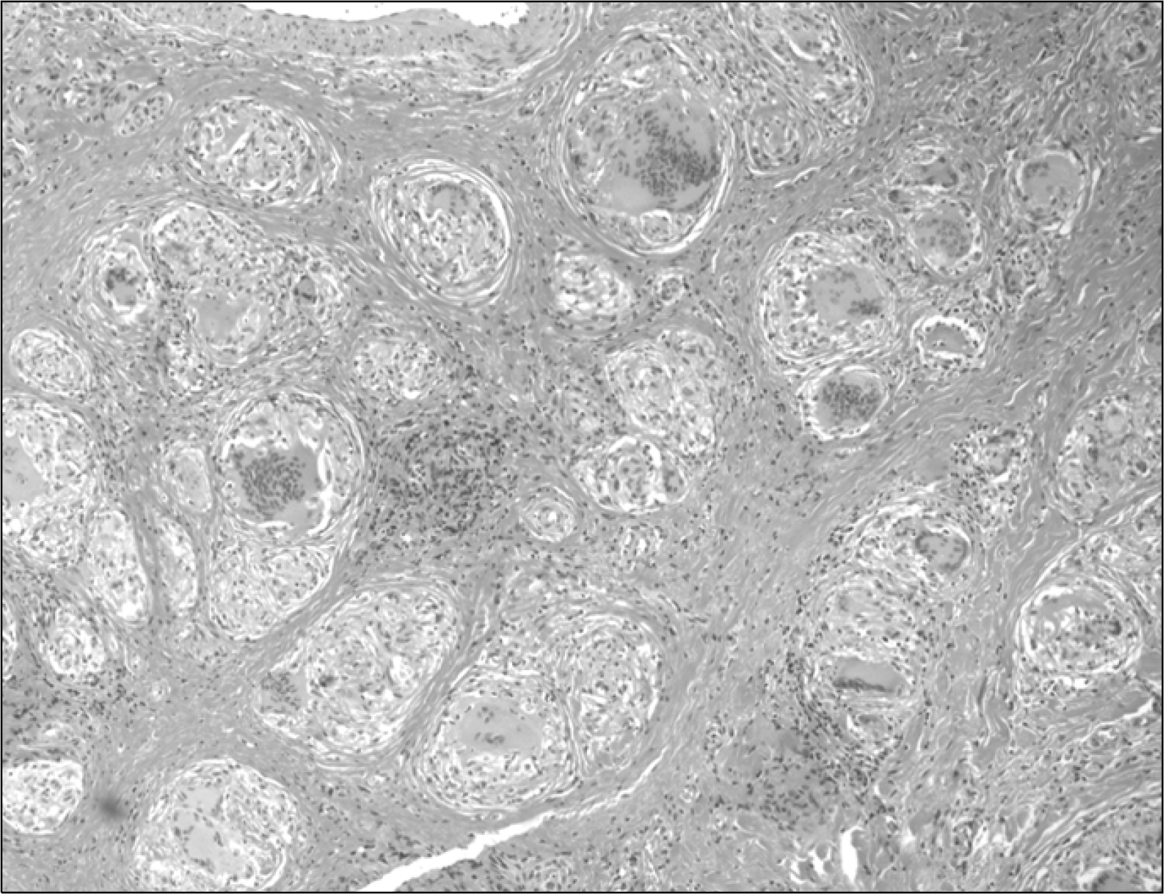
Sarcoidosis is multi-systemic disorder of an unknown etiology, and this is histologically characterized by noncaseating granulomatous inflammation. Sarcoidosis may affect the lung, skin, lymph nodes and eyes, but it rarely affects the subcutaneous tissue. There has been no report of diffuse subcutaneous sarcoidosis in Korea. We experienced a 57-year-old female with diffuse subcutaneous sarcoidosis that presented as thickened extremities. The patient complained of edema and skin thickening on both upper extremities. Magnetic resonance imaging revealed the reticular form of sarcoidosis on the forearm and the biopsy showed noncaseating granuloma. She was finally diagnosed as diffuse subcutaneous sarcoidosis and she improved after treatment with corticosteroid. We report here on this unusual case along with a review of the relevant literature.
Go to : 
REFERENCES
2). Imboden J., Hellmann DB., Stone JH. Current rheumatology diagnosis & treatment. 1st ed.p. 195. U.S.A.: McGraw Hill;2004.
3). Choi KH., Choi YS., Kim BS., Joo JE., Jung YY., Cho YK, et al. A nodular type of subcutaneous sarcoidosis. J Korean Soc Radiol. 2009. 60:47–50.
4). Lynch 3rd JP., White ES. Pulmonology sarcoidosis. Eur Restpir Mon. 2005. 32:105.
7). Hong YC., Na DJ., Han SH., Lee YD., Cho YS., Han MS, et al. A case of scar sarcoidosis. Korean J Intern Med. 2008. 23:213–5.

9). Kim JP., Kim SH., Lee HG., Sung MS., Kim YS., Kim HM, et al. Muscle mass in the calf as a presenting symptom of sarcoidosis. J Korean Rheum Assoc. 2003. 10:66–70.
10). Behbenani N., Jayakrishnan B., Khadadah M., Al-Sawi M. Long term prognosis of sarcoidosis in Arabs and Asians: predictors of good outcome. Sarcoidosis Vasc Diffuse Lung Dis. 2005. 23:209–14.
11). Do YS., Lee JY., Kim HJ., Kim EH., Chai IY., Jeon CH, et al. A case of sarcoidosis presented with myofasciitis. J Korean Rheum Assoc. 2005. 12:42–6.
13). Baughman RP., Lower EE. Newer therapies for cutaneous sarcoidosis: the role of thalidomide and other agents. Am J Clin Dermatol. 2004. 5:285–94.
14). Lee MJ., Lew W., Lee SH. A case of psoriasiform sarcoidosis as early manifestation of systemic sarcoidosis. Korean J Dermatol. 2004. 42:1606–9.
Go to : 
 | Fig. 1.The sagittal (A) and axial sections (B) of the Gd-DTPA enhanced T1-weighted magnetic resonance images. These images show the reticular pattern with low to intermediate signal intensity (arrows). |
 | Fig. 2.The skin biopsy specimen shows many noncasating granulomas that consisted of epithelial cells, multinucleated giant cells and lymphocytes (H&E stain, ×100). |
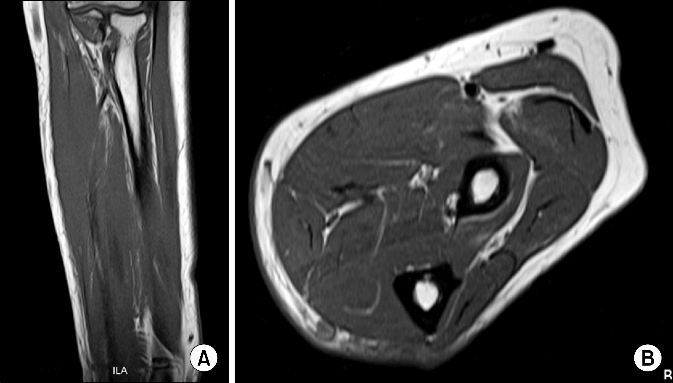
 | Fig. 3.The sagittal (A) and axial sections (B) of the Gd-DTPA enhanced T1-weighted magnetic resonance images. These images show disappearance of the reticular pattern and the decreased extent of the high signal intensity of the medial subcutaneous fat layer in the left forearm. |
Table 1.
Cases of soft tissue sarcoidosis in Korea


 PDF
PDF ePub
ePub Citation
 Print
Print


 XML Download