REFERENCES
2). Keith MW., Masear V., Chung K., Maupin K., Andary M., Amadio PC, et al. Diagnosis of carpal tunnel syndrome. J Am Acad Orthop Surg. 2009. 17:389–96.

3). Bianchi S., Montet X., Martinoli C., Bonvin F., Fasel J. High-resolution sonography of compressive neuropathies of the wrist. J Clin Ultrasound. 2004. 32:451–61.

4). SarriAa L., Cabada T., Cozcolluela R., MartiAnez-Berganza T., GarciAa S. Carpal tunnel syndrome: usefulness of sonography. Eur Radiol. 2000. 10:1920–5.
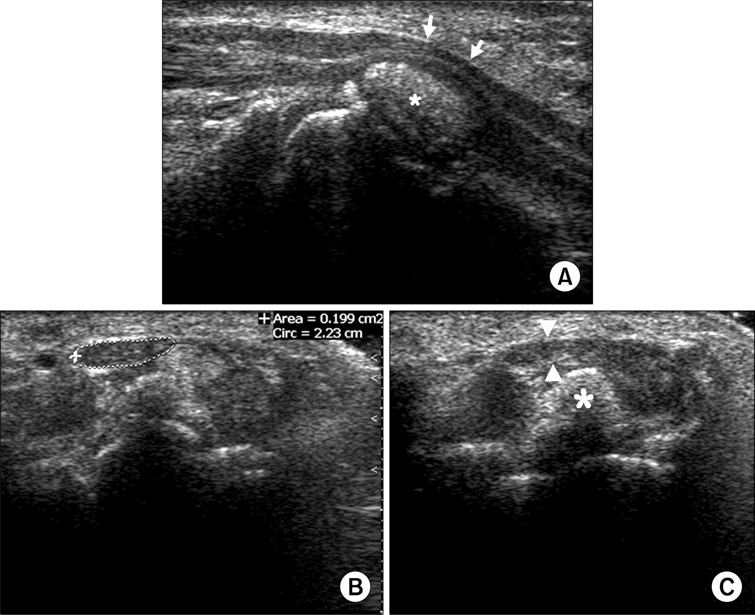
Fig. 1.
Ultrasonography shows (A) 12×6 mm sized oval shaped hyperechoic mass (∗) in the carpal tunnel compressing the median nerve (arrows) in longitudinal scan, (B) increased cross-sectional area of medial nerve in the tunnel inlet, and (C) the flattening of median nerve (arrow heads) near the hyperechoic mass (∗) in mid-tunnel in transverse scans.


 PDF
PDF ePub
ePub Citation
 Print
Print


 XML Download