Abstract
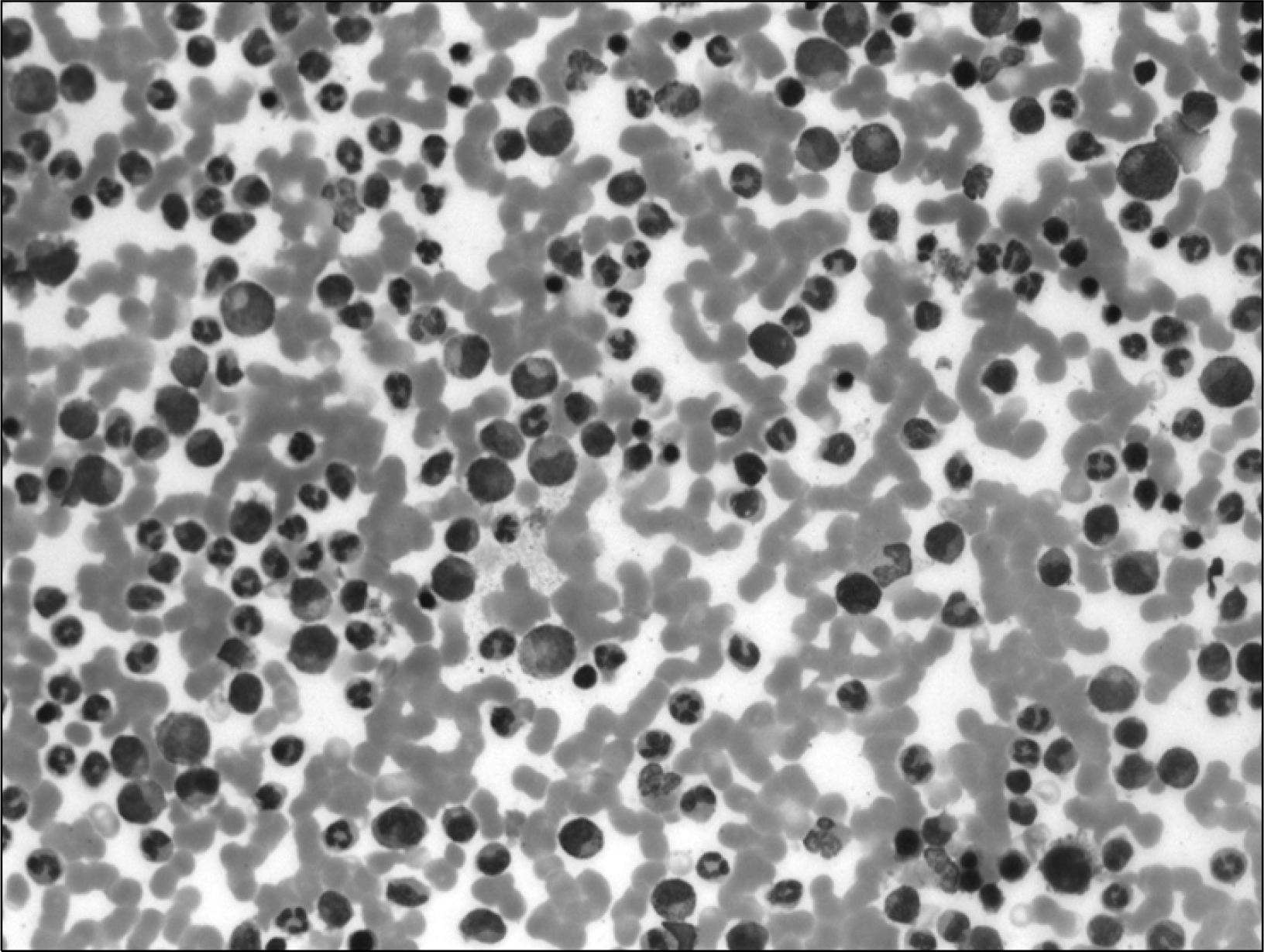
Ankylosing spondylitis (AS) is occasionally accompanied by hematological malignancies such as myelodysplastic syndrome, acute myelogenous leukemia, or multiple myeloma. Chronic myelogenous leukemia (CML) is a myeloproliferative disorder associated with Philadelphia chromosome and is usually treated with imatinib, which inhibits tyrosine kinases. Although there have been reports of CML cases accompanied by several rheumatic diseases such as rheumatoid arthritis, Behcet's disease, systemic sclerosis, or undifferentiated spondylopathy, no studies have reported a case of CML with AS. We experienced a 50-year-old male patient who presented with buttock and low back pain and was diagnosed with both AS and CML. Magnetic resonance imaging showed sacroiliitis along with abnormal marrow infiltration, and a bone marrow biopsy confirmed the CML diagnosis. He was treated with imatinib, which was effective for the CML but not for the AS. This is the first case report of AS accompanied by CML.
REFERENCES
1). Yang CH., Jeong MK., Lee HJ., Lee YH., Yoon KY., Kim JS, et al. Multiple myeloma combined with ankylosing spondylitis. Korean J Intern Med. 1985. 28:560–6.
2). Kim YN., Lee HE., Lee SH., Lee YA., Woo DH., Hwangbo Y, et al. Ankylosing spondylitis associated with plasmacytoma. J Korean Rheum Assoc. 2005. 12:240–4.
3). Lee JJ., Kim HG., Ahn JK., Hwang JW., Jang JH., Koh EM, et al. Ankylosing spondylitis in a patient with myelodysplatic syndrome: an association with HLA-B27 or coincidence? Rheumatol Int. 2009. 29:689–92.
4). Hoshino T., Matsushima T., Saitoh Y., Yamane A., Takizawa M., Irisawa H, et al. Sacroiliitis as an initial manifestation of acute myelogenous leukemia. Int J Hematol. 2006. 84:421–4.

5). Au WY., Hawkins BR., Cheng N., Lie AK., Liang R., Kwong YL. Risk of haematological malignancies in HLA-B27 carriers. Br J Haematol. 2001. 115:320–2.

6). Senel S., Kaya E., Aydogdu I., Erkurt MA., Kuku I. Rheumatic diseases and chronic myelogenous leukemia, presentation of four cases and review of the literature. Rheumatol Int. 2006. 26:857–61.

7). Miyachi K., Ihara A., Hankins RW., Murai R., Machiro S., Miyashita H. Efficacy of imatinib mesylate (STI571) treatment for a patient with rheumatoid arthritis developing chronic myelogenous leukemia. Clin Rheumatol. 2003. 22:329–32.

8). Sacchi S., Kantarjian H., O'Brien S., Cohen PR., Pierce S., Talpaz M. Immune-mediated and unusual complications during interferon alfa therapy in chronic myelogenous leukemia. J Clin Oncol. 1995. 13:2401–7.

9). Nobauer I., Uffmann M. Differential diagnosis of focal and diffuse neoplastic diseases of bone marrow in MRI. Eur J Radiol. 2005. 55:2–32.
10). Paniagua RT., Robinson WH. Imatinib for the treatment of rheumatic diseases. Nat Clin Pract Rheumatol. 2007. 3:190–1.

11). Eklund KK., Joensuu H. Treatment of rheumatoid arthritis with imatinib mesylate: clinical improvement in three refractory cases. Ann Med. 2003. 35:362–7.

12). Nazarinia MA., Ghaffarpasand F., Seraj SR., Heiran HR., Khojasteh HN. Imatinib treatment of ankylosing spondylitis in a patient resistant to NSAIDs and Infliximab. J Clin Rheumatol. 2010. 16:140–2.

13). Bakland G., Nossent H. Acute myelogenous leukaemia following etanercept therapy. Rheumatology. 2003. 42:900–1.

14). Nair B., Raval G., Mehta P. TNF-aL inhibitor etanercept and hematologic malignancies: report of a case and review of the literature. Am J Hematol. 2007. 82:1022–4.
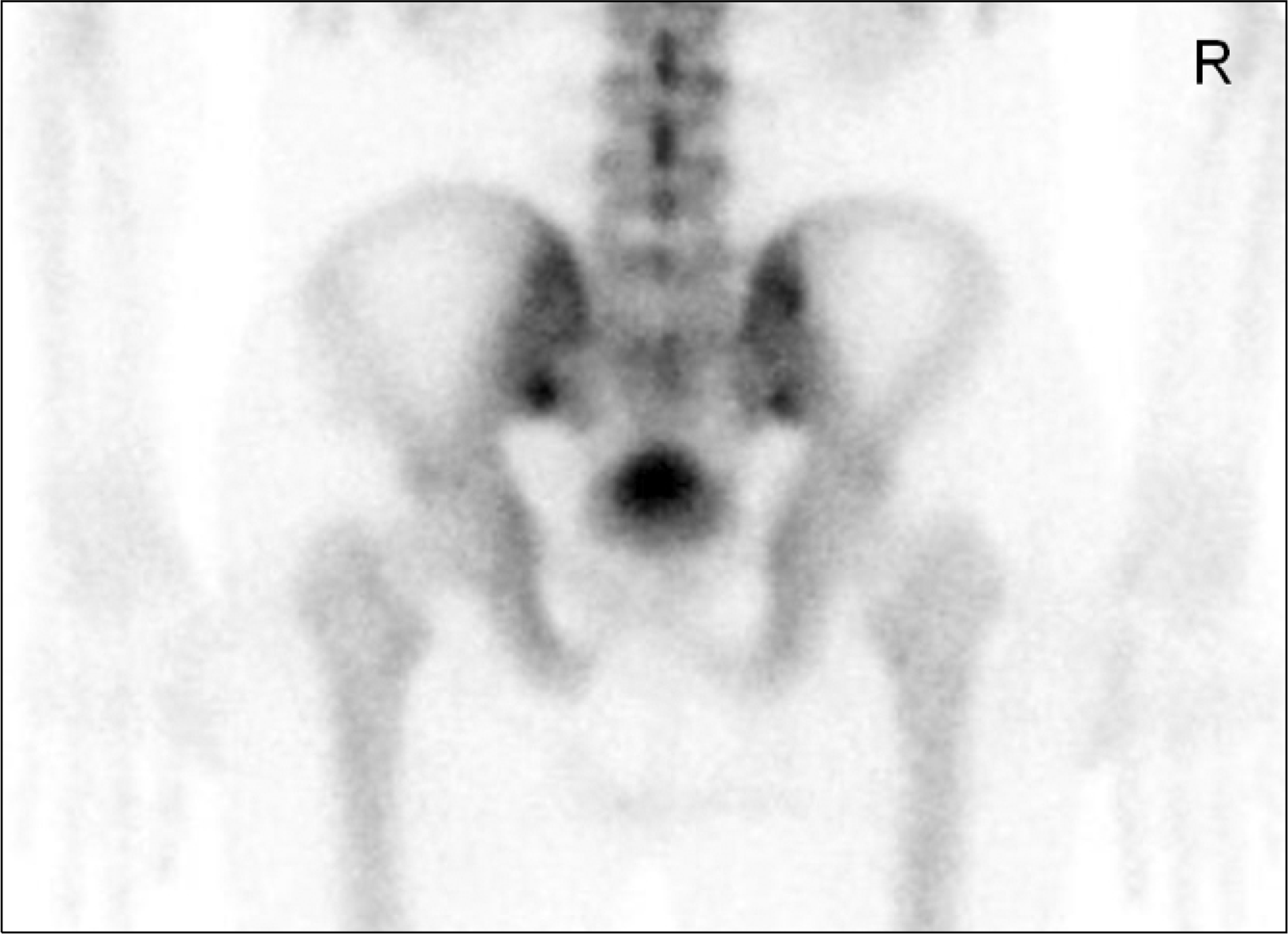
Fig. 1.
Bone scan (posterior view of the pelvis) shows increased radio-uptakes in both sacroiliac joints.

Fig. 2.
(A) T2-weighted fat suppression magnetic imaging shows high signal intensity in both sacroiliac joints (black arrows) and bone marrow edema in the left sacroiliac joint. Additionally, abnormal bone marrow infiltrations are present in the femurs (white arrows) and the pelvic bones. (B) T1-weighted imaging clearly shows abnormal bone marrow infiltrations in the femur (arrows) and the pelvic bones.


 PDF
PDF ePub
ePub Citation
 Print
Print


 XML Download