

 PDF
PDF ePub
ePub Citation
 Print
Print


Abstract
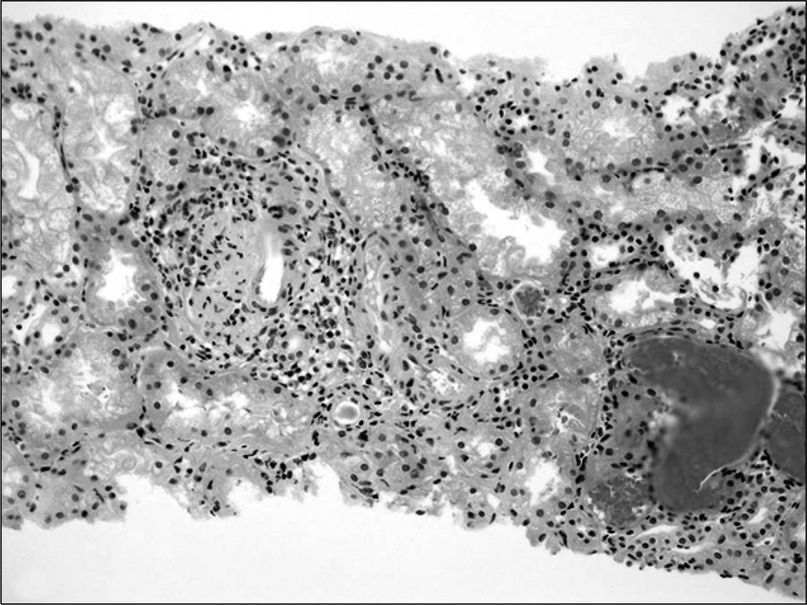
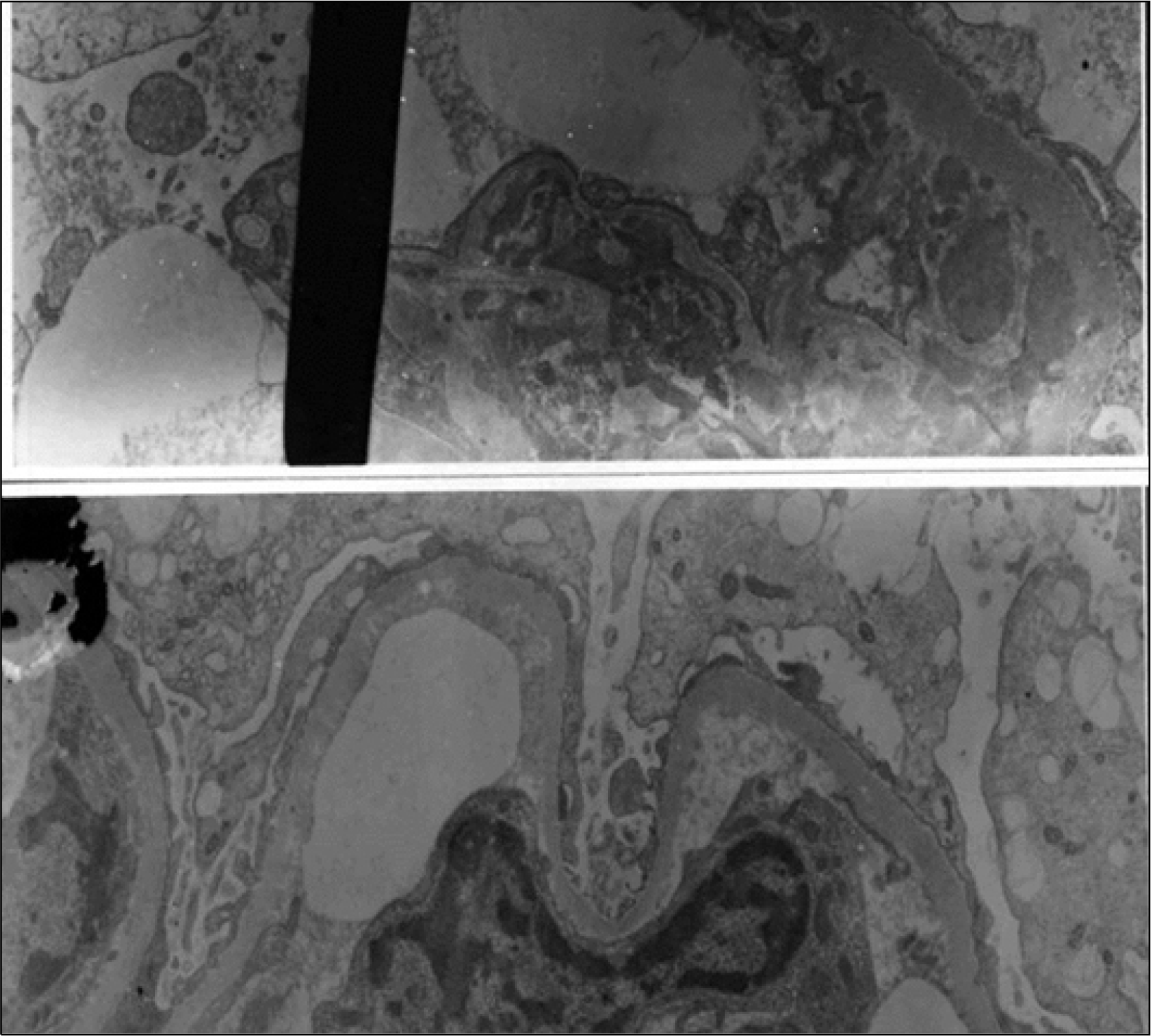
Microscopic polyangiitis (MPA) is characterized by pauci-immune necrotizing small vessel vasculitis without clinical or pathological evidence of necrotizing granulomatous inflammation. The kidney is the most often affected organ in the majority of patients with MPA, and renal manifestations are usually the first symptoms. Glomerular capillaries are affected most often, resulting in necrotizing glomerulonephritis, usually in a crescent formation, with no or few immune deposits able to be demonstrated at the sites of vasculitis and glomerulonephritis. We report a case of microscopic polyangiitis in both legs with pitting edema in a 50-year-old female. Laboratory findings showed hematuria, proteinuria, and a positive peripheral antineutrophil cytoplasmic antibody. A renal biopsy revealed pauci-immune splitting and necrotizing capillary loop walls necrotizing vasculitis and membranoproliferative glomerulonephritis (MPGN). With a diagnosis of MPA, she has been managed with high dose steroid and cyclophosphamide. To our knowledge, this is the first reported case of MPA with MPGN.
REFERENCES
1). Davson J., Ball J., Platt R. The kidney in periarteritis nodosa. Q J Med. 1948. 17:175–92.
2). Jennette JC., Falk RJ., Andrassy K., Bacon PA., Churg J., Gross WL, et al. Nomenclature of systemic vasculitides. Proposal of an international consensus conference. Arthritis Rheum. 1994. 37:187–92.
3). Lhote F., Cohen P., Guillevin L. Polyarteritis nodosa, microscopic polyangiitis and Churg-Strauss syndrome. Lupus. 1998. 7:238–58.

4). Valente RM., Conn DL. Polyarteritis-Polyarteritis nodosa and microscopic polyangiitis. In:. Kippel JH, Dieppe PA, editors. eds.Rheumatology. 2nd ed.p. 201–11. London: Mosby;1998.
5). Gaskin G., Pusey C. Systemic vasculitis. In:. Davison AM, Cameron JS, Grunfeld JP, Kerr DM, Ritz E, Winearls CG, editors. eds.Oxford textbook of clinical nephrology. 2nd ed.p. 877–901. Oxford, Oxford University Press;1998.
6). Seo CG., Lee SH., Kim SH., Kim KC., Kim MS., Han SB, et al. A case of microscopic polyangiitis presenting as diffuse alveolar hemorrhage. Tuberc Respir Dis. 2002. 53:202–8.
7). Savage CO., Winearls CG., Evans DJ., Rees AJ., Lockwood CM. Microscopic polyarteritis: presentation, pathology and prognosis. Q J Med. 1985. 56:467–83.
8). Guillevin L., Durand-Gasselin B., Cevallos R., Garyraud M., Lhote F., Callard P, et al. Microscopic polyangiitis: clinical symptoms, laboratory findings and outcome in 89 patients. Arthritis Rheum. 1997. ;40 (Suppl): S169.
9). Balow JE., Fauci AS. Vasculitic disease of the kidney. In:. Schreier RW, Cottschalk CW, editors. eds.Disease of the kidney. 6th ed.p. 1851–67. New York, Little Brown & Company;1997.
10). Gaber LW., Wall BM., Cooke CR. Coexistance of antineutrophil cytoplasmic antibody-associated glomerulonephritis and membranous glomrulopathy. Am J Clin Pathol. 1993. 99:211–5.
11). Tse WY., Howie Adu D., Savage COS., Richards NT., Wheeler DC., Michael J. Association of vasculitic glomerulonephritis with membranous nephropathy: a report of 10 cases. Nephrol Dial Transplant. 1997. 12:1017–27.
12). Green RJ., Ruoss SJ., Kraft SA., Duncan SR., Berry GJ., Raffin TA. Pulmonary capillaritis and alveolar hemorrhage: update on diagnosis and management. Chest. 1996. 110:1305–16.
13). Guillevin L., Lhote F. Treatment of polyarteritis nodosa and microscopic polyangiitis. Arthritis Rheum. 1998. 41:2100–5.
14). Hogan SL., Nachman PH., Wikman AS., Jennette JC., Falk RJ. Prognostic markers in patients with anti-neutrophil cytoplasmic autoantibody-associated microscopic polyangiitis and glomerulonephritis. J Am Soc Nephrol. 1996. 7:23–32.
15). Jennette JC., Wilkman AS., Falk RJ. Diagnostic predictive value of ANCA serology. Kidney Int. 1998. 53:796–8.
 XML Download