Abstract
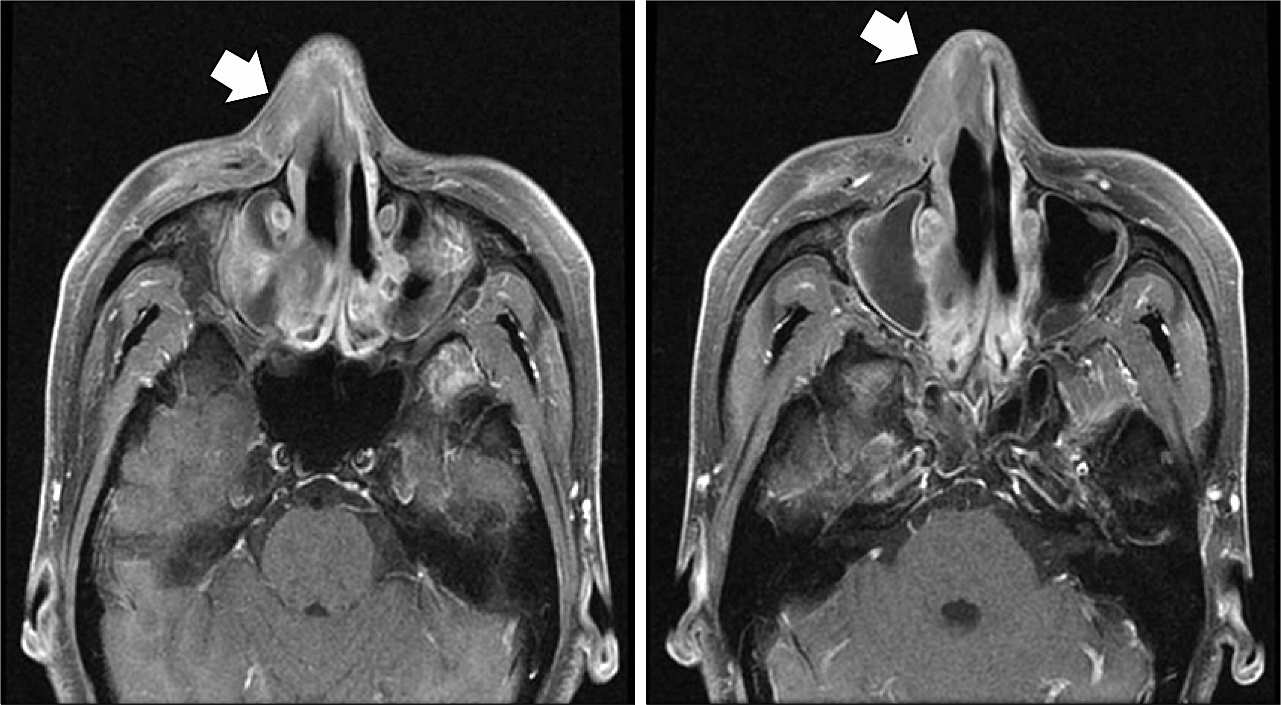
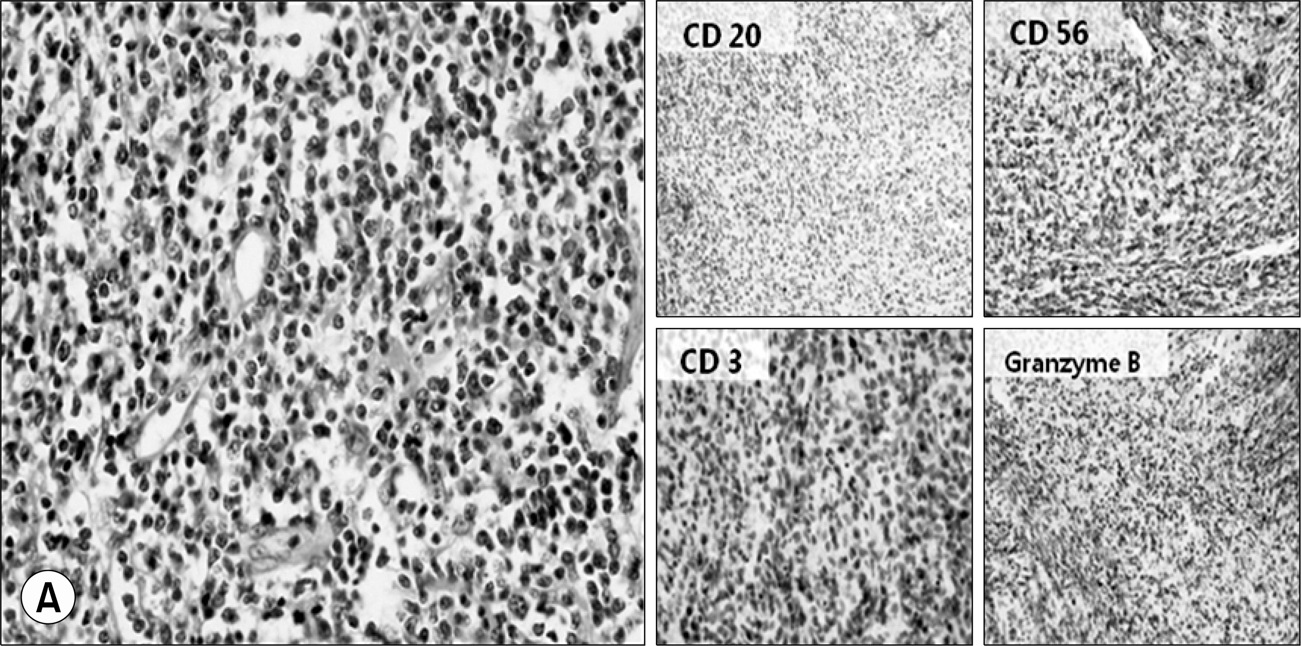
It is occasionally difficult to distinguish Wegener's granulomatosis (WG) from other diseases including malignancy, tuberculosis, and various types of vasculitis because of the overlapping symptoms and signs. We report on a patient with NK/T cell lymphoma who was treated with a limited form of WG. At his first visit, he presented with left foot drop and recurrent nasal swelling. Necrosis and massive infiltration of inflammatory cells were identified on a nasal tissue biopsy. Sural nerve biopsy findings also showed infiltration of inflammatory cells in both the endoneurium and perivascular area; thus, a diagnosis of a limited form of WG was made. After combination therapy with a glucocorticoid and oral cyclophosphamide was initiated, his condition completely recovered without recurrence for the next 2 years. However, he visited the hospital again for recurrence of nasal swelling. Repeated biopsy of nasal tissues, combined with an immunophenotypic analysis revealed NK/T cell lymphoma. The possibility of NK/T lymphoma should be considered when evaluating a limited type of WG, which shows atypical findings on biopsy as well as recurrent deterioration, as a suboptimal dose of immunosuppressive therapy may mask its expression and lead to a poor prognosis.
REFERENCES
1). Thajeb P., Tsai JJ. Cerebral and oculorhinal manifestations of a limited form of Wegener's granulomatosis with c-ANCA-associated vasculitis. J Neuroimaging. 2001. 11:59–63.

2). Cassan SM., Coles DT., Harrison EG Jr. The concept of limited forms of Wegener's granulomatosis. Am J Med. 1970. 49:366–79.

3). Hoffman GS., Kerr GS., Leavitt RY., Hallahan CW., Lebovics RS., Travis WD, et al. Wegener granulomatosis: an analysis of 158 patients. Ann Intern Med. 1992. 116:488–98.

4). Knight A., Askling J., Ekbom A. Cancer incidence in a population-based cohort of patients with Wegener's granulomatosis. Int J Cancer. 2002. 100:82–5.

5). Fain O., Hamidou M., Cacoub P., Godeau B., Wechsler B., Paries J, et al. Vasculitides associated with malignancies: analysis of sixty patients. Arthritis Rheum. 2007. 57:1473–80.

6). Manske CL., Glick AD., Stone WJ. Sinusitis and glomerulonephritis in a middle-aged man. Am J Kidney Dis. 1987. 10:320–2.

7). Borress RS., Maccabee P., Har-El G. Foot drop in head and neck cancer. Am J Otolaryngol. 2007. 28:321–4.

8). VaAroAczy L., Gergely L., Zeher M., Szegedi G., IlleAs A. Malignant lymphoma-associated autoimmune diseases- a descriptive epidemiological study. Rheumatol Int. 2002. 22:233–7.
Fig. 1.
Initial radiologic examinations and tissue biopsy. (A) Paranasal sinusoid computed tomography scan on admission (3 years ago) shows thickening of the mucoperiosteal lining of the frontal sinus, ethmoid sinus, and maxillary sinuses. Near total opacification of the right maxillary sinus and mucosal thickening of the left maxillary sinus are also seen. (B) A focally dense, mass-like lesion was seen in the anterior nostril area. (arrow) (C) Necrosis and massive infiltration of inflammatory cells were observed on a nasal tissue biopsy. (D) A sural nerve biopsy indicated that inflammatory cells had infiltrated both the endoneurium with axonal degeneration and the perivascular area.


 PDF
PDF ePub
ePub Citation
 Print
Print


 XML Download