Journal List > J Korean Rheum Assoc > v.17(1) > 1003731

Oh, Park, Seo, and Park: Meta-analysis on the Risk Factors for Fracture in Patients with Rheumatoid Arthritis

Abstract

Objective

This study was conducted by using a meta-analysis to calculate the mean effect sizes of the risk factors for fracture of patients with rheumatoid arthritis (RA) and to perform tests for the significance of the calculated mean effect sizes.

Methods

Eleven studies that directly examined the relationships between fracture and risk factors were selected from 179 related studies identified from PubMed, MEDLINE, COCHRAN and CINHAL with the key words being ‘rheumatoid arthritis’, ‘fracture’ and ‘risk factors’.

Results

The mean effect size of age on fracture was significant. The mean effect sizes of steroid use and the duration of RA on fracture were also significant. However, all of these effect sizes (age, the use of steroid and the duration of disease) were small (bar D=0.15, 0.16 and 0.12, respectively). The mean effect size of the BMD at the lumbar spine was significant and the effect of the BMD at the L-spine was of medium size (bar D=0.47). The mean effect sizes of the other risk factors such as disability, disease activity, the BMD at the femur neck, BMI and a smoking habit were not significant.

Conclusion

From the study results, it was noted that the efforts to prevent fracture of RA patients should focus on patients with an older age, the patients who used steroid, the patients with a longer duration of RA and the patients who present with low BMD at the lumbar spine.

REFERENCES

1). Spector TD., Hall GM., McCloskey EV., Kanis JA. Risk of vertebral fracture in women with rheumatoid arthritis. BMJ. 1993. 306:558.
crossref
2). Kim JY., Lee YW., Ham OK. Factors related to fall in elderly patients with osteoprosis. J Korean Acad Adult Nurs. 2009. 21:257–67.
3). Oh HY., Im YM. Functional status and health care utilization among elders with hip fracture surgery from fall. J Korean Acad Adult Nurs. 2003. 15:432–40.
4). Kay LJ., Holland TM., Platt PN. Stress fractures in rheumatoid arthritis: a cross series and case-control study. Ann Rheum Dis. 2004. 63:1690–2.
5). van Staa TP., Geusens P., Bijlsman JW., Leufkens HG., Cooper C. Clinical assessment of the long-term risk of fracture in patients with rheumatoid arthritis. Arthritis Rheum. 2006. 54:3104–12.
crossref
6). Furuya T., Kotake S., Inoue E., Nanke Y., Yago T., Kobashigawa T, et al. Risk factors asociated with incident clinical vertebral and nonvertebral fractures in Japanese women with rheumatoid arthritis: a prospective 54-month obsevational study. J Rheumatol. 2007. 34:303–10.
7). Arai K., Hanyn T., Sugitani H., Murai T., Fujisawa J., Nakazono K, et al. Risk factors for vertebral fracture in menopausal or post menopausal Japanese women with rheumatoid arthritis: a cross-sectional and longitudinal study, J Bone Miner Metab. 2006. 24:118–24.
8). de Nijs RNJ., Jacobs JWG., Bijlsma JWJ., Lems WF., Laan RFJM., Houben HHM, et al. On behalf of the Osteoporosis Working Group of the Dutch Society of Rheumatology Prevalence of vertebral deformities and symptomatic vertebral fractures in corticosteroid treated patients with rheumatoid arthritis. Rheumatology. 2001. 40:1375–83.
9). PeArez-Edo L., Diez-PeArez A., MarinToso L., ValleAs A., Serrano S., Carbonell J. Bone metabolism and histomorphometric changes in rheumatoid arthritis. Scand J Rheumatol. 2002. 31:285–90.
10). Adachi JD., Ioannidis G., Pickard L., Berger C., Prior JC., Joseph L, et al. The association between osteoporotic fractures and health-related quality of life as measured by the Health Utilities Index in the Canadian Multicentre Osteoporosis Study (CaMos). Osteoporos Int. 2003. 14:895–904.
crossref
11). Lee HY., Bak WS. The risk factors of osteoporosis in Korean postmenopausal women. J Korean Acad Adult Nurs. 2009. 21:303–13.
12). Irvine DM., Vincent L., Graydon JE., Bubela N. Fatigue in women with breast cancer receiving radiation therapy. Cancer Nurs. 1998. 21:127–35.
crossref
13). Song HH. Meta-Analysis for Researches in Medical, Nursing, and Social Science. Seoul, Chung-Moon Gak. 1998.
14). Kim NC., Song HH., Kim JO. Effects of nursing interventions applied to surgery patients: a meta-analysis. J Korean Acad Adult Nurs. 1998. 10:523–34.
15). Fujiwara S., Nakamura T., Orimo H., Hosoi T., Gorai I., Oden A, et al. Development and application of a Japanese model of WHO fracture risk assessment tool (FRAXTM). Osteoporos Int. 2008. 19:429–35.
16). Gronowicz G., McCarthy MB. Glucocorticoids inhibit the attachment of osteoblasts to bone extracellular matrix proteins and disease bT-1 integrin levels. Endocrinology. 1995. 136:598–608.
17). Verstraeten A., Dequeker J. Vertebral and peripheral bone mineral content and fracture incidence in postmenopausal patients with rheumatoid arthritis: effect of low dose corticosteroids. Ann Rheum Dis. 1986. 45:852–7.
crossref
18). Laan RF., van Riel PL., van Erning LJ., LEmmens JA., Rujs SH., van de Putte LB. Vertebral osteoporosis in rheumatoid arthritis patients: effect of low dose prednisone therapy. Br J Rheumatol. 1992. 31:91–6.
crossref
19). Butler RC., Davie MW., Worsfold M., Sharp CA. Bone mineral content in patients with rheumatoid arthritis: Relationship to low-dose steroid therapy. Br J Rheumatol. 1991. 30:86–90.
crossref
20). Sambrook PN., Cohen ML., Eisman JA., Pocock NA., Eberl S., Champion GD, et al. Effects of low dose corticosteroids on bone mass in rheumatoid arthritis: a longitudinal study. Ann Rheum Dis. 1989. 48:535–8.
crossref
21). Leboff MS., Wade JP., Mackowiak S., Fuleihan ., Zangari M., Liang MH. Low dose prednisonlon does not affect calcium homeostasis or bone dinsity in postmenopausal women with rheumatoid arthriti. J Rheumatol. 1991. 18:330–44.
22). Michel BA., Bloch DA., Wolfe F., Fries JF. Fracture in rheumatoid arthritis: an evaluation of associated risk factors. J Rheumatol. 1993. 20:1666–9.
23). Paganini-Hill A., Ross RK., Gerkins VR., Henderson BE., Arthur M., Mack TM. Menopausal estrogen therapy and hip fracture. Ann Int Med. 1981. 95:28–31.
24). Michel BA., Bloch DA., Fries JF. Predictors of fractures in early rheumatiod arthritis. J Rheumatol. 1991. 18:804–8.
25). Arend WP., Dayer JM. Cytokines and cytokine inhibitors or antagonists in rheumatoid arthritis. Arthritis Rheum. 1990. 33:305–15.
crossref
26). Dequecker J., Geusens P. Osteoporosis and Arthritis. Ann Rheum Dis. 1990. 49:276–80.
27). Peel NF., Moore DJ., Barrington NA., Bax DE., Eastell R. Risk of vertebral fracture and relationship to bone mineral density in steroid treated rheumatoid arthritis. Ann Rheum Dis. 1995. 54:801–6.
crossref
28). Nampei A., Hashimoto J., Koyanagi J., Ono T., Hashimoto H., Tsumaki N, et al. Characteristics of fracture and related factors in patients with rheumaotid arthritis. Mod Rheumatol. 2008. 18:170–6.
29). Coulson K., Reed G., Gilliam BE., Kremer JM., Pepmueller PH. Factors influencing fracture risk, T score, and management of osteoporosis inpatients with rheumatoid arthritis in the consortium of rheumatology researchers of north america (CORRONA) registry. J Clin Rheumatol. 2009. 15:155–60.
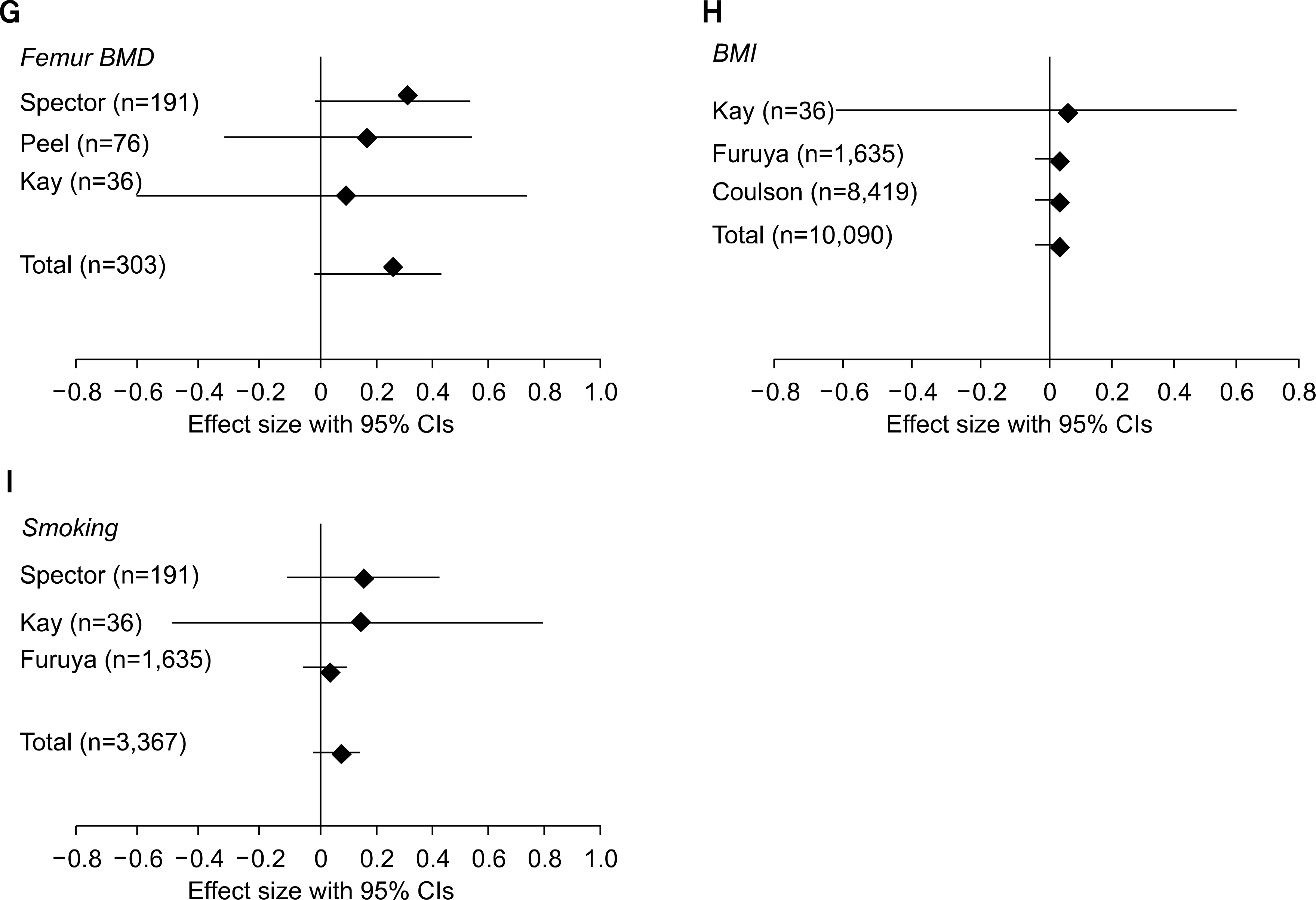
Fig. 1.
Effect size of individual study for risk factors on fracture of rheumatoid arthritis patients. (A) Effect size of age on fracture, (B) Effect size of steroid on fracture, (C) Effect size of disability on fracture, (D) Effect size of duration on fracture, (E) Effect size of disease activity on fracture, (F) Effect size of L-spine BMD on fracture, (G) Effect of Femur BMD on fracture, (H) Effect of BMI on fracture, (I) Effect of smoking on fracture.
jkra-17-23f1a.tifjkra-17-23f1b.tif
Table 1.
The sample size, sample characteristics, fracture site, BMD site, and results, and the quality score of the reviewed studies
Author 1. Sample size
2. Subjects' characteristics
3. Study design∗
1. Fracture site
2. BMD sites
1. % of fracture patients
2. Risk factors and statistics
Quality score
1. Spector, et al. (1) 1. 191
2. Menopaused RA women
1. Spine fx
2. Femur neck, L1-4
1. 12.1%
2. 1) Age: p=0.02, 2) Duration of disease: p=0.37, 3) ESR: t=-0,10, 4) HAQ: t=0.04, 5) Disability: p=0.14, 6) Duration of disease: t=0.99, 7) Menopaused age: p=0.60, 8) BMI: p=0.41, 9) Use of steroid: Z=–0.58, 10) BMD: L (t=–0.6), femur (t=–1.75), 11) Smoking: p=0.04
4
2. Peel, et al. (27) 1. 76
2. Menopaused RA women (duration: 90%, 15 years; steroid: 85%, > 10 years)
1. Vertebral fx
2. Lumbar, femur
1. 27.6% (vertebral deformity)
2. 1) L: t=–0.75, 2) Femur: t=–0.5
4
3. de Nijs, et al. (8) 1. 500
2. RA patients
1. Spine
2. –
1. 19.0%
2. 1) Use of steroid: Z=3.0 (vetebral fracture)
4
4. Perez-Edo, et al. (9) 1. 66
2. RA patients
1. Spine fx
2. Radius, Lumbar
1. 30%
2. Age: p=0.04
2
5. Arai, et al. (7) 1. 117
2. Menopaused RA women (Japan)
1. Vertebral fx
2. Lumbar (L2-L4)
1. 21%
2. 1) Use of steroid: Z=2.75, 2) BMD: Z=3.91, 3) Age: Z=3.5
3
6. Kay, et al. (4) 1. 36
2. RA patients
1. Stress fx
2. Femur, lumbar
1. 0.8%
2. 1) Age: p=0.41, 2) Duration of disease: p=0.37, 3) RF: p=0.18, 4) Disability: p=0.14, 5) Arthroplasty: p=0.73, 6) Menopaused age: p=0.60, 7) BMI: p=0.41, 8) Use of steroid: p=0.32, 9) BMD: Lumbar (p=0.77), femur (p=0.59), 10) Smoking: p=0.81
2
7. Michel, et al. (24) 1. 395
2. RA patients (mean age: 49 years, duration: 9.3 years)
1. Fracture all over the body
2. –
1. 13.2%
2. 1) Steroid: p=0.026, 2) Duration: p=0.083
4
8. Furuya, et al. (6) 1. 1,635
2. RA women above 50 years old
1. Fracture all over the body (spine, wrist, hip, pelvis, ribs)
2. –
1. 17.6%
2. Selected only vertebral fracture 1) Age: t=–1.08, 2) BMI: t=0.11, 3) Smoking: Z=0.33, 4) Alcohol: Z=–0.83, 5) Duration of disease: t=–0.80, 6) RF: t=–0.53, 7) ESR: t=–0.64, 8) Pain: t=–0.74, 9) HAQ-J: t=–1.52, 10) Prior fx: Z=1.25, 11) Orthopedic surgery: Z=0.44, 12) Use of steroid: Z=2.44
4
9. Michel, et al. (22) 1. 1,110
2. RA patients (mean age: 54, mean duration of disease: 8.4 years)
1. Fracture all over the body
2. –
1. 20.4%
2. 1) Duration of steroid use: Z=6.0, 2) Grip power: Z=3.67, 3) Disability: Z=5.0, 4) Duration: Z=3.75, 5) BMI: Z=4.58, 6) Age: Z=4.4
4
10. Nampei, et al. (28) 1. 209
2. RA outpatients
1. Vertebral+non vertebral+insufficiency fx
2. –
1. 11.5%
2. Only vertebra fx (multivariate analysis)
1) Steroid: p=0.032
3
11. Coulson, et al. (29) 1. 8,419
2. RA female patients
1. Spine, hip, nonspine (wrist, ribs, pelvis, other)
2. L-spine, femur
1. 431/8,419(5.1%) subjects with fracture (spine: 99, hip: 77, others: 326)
2. Spine fracture
1) Postmenopausal: p=0.15, 2) BMI: p=0.38, 3) Marial status: p=0.86, 4) HAQ: p=0.77, 5) Steroid: 0.52, 6) TNF: p=0.08, 7) TNF+MTX: p=0.51, 8) DMARDS: p=0.35, 9) MTX-DMARDS: p=0.62
3

∗A11 of the studies included in this meta-analysis adopted a non-experimental correlational research design, If Z or t ≥ 1.96, p≤0.05; if Z or t≥2.54, p≤0.01

Table 2.
The results of the meta-analyses for the fracture risk factors of rheumatoid arthritis patients
Domain Parameters n $DM∗(95% CI) Sig. U (p) $RM Homogeneity§ Q (p) nfs||
Age 6 0.15 (0.08 ~ 0.22) 17.68 (<0.001) 0.08 4.53 (0.211) 2.18
Medication Use of steroid treatment 7 0.16 (0.08 ~ 0.24)∗∗ 16.24 (<0.001) 0.08 10.83 (0.060) 1.23
Disability Health assessment Questionnaire-disability index 6 0.03 (–0.007 ~ 0.007) 2.65 (0.100) 0.02 5.54 (0.242) 4.21
Duration of disease 5 0.12 (0.05 ~ 0.18) 11.76 (0.011) 0.06 6.06 (0.200) 2.08
Disease activity ESR (mm/hr), Rheumatoid factor (U/ml) 4 0.03 (–0.03 ~ 0.10) 0.83 (0.362) 0.02 0.67 (0.881) 3.39
Bone mineral Lumbar spine BMD (%) 4 0.47 (0.21 ~ 0.74)†† 12.60 (<0.001) 0.23 4.57 (0.102) 4.17
Density Femur BMD (%) 3 0.20 (–0.03 ~ 0.42) 2.92 (0.092) 0.10 0.42 (0.813) 1.00
Body weight Body mass index (kg/m2) 4 0.01 (–0.03 ~ 0.05) 0.11 (0.741) 0.003 0.04 (0.982) 2.90
Health habit Smoking status (smoker vs non smoker) 3 0.04 (–0.05 ~ 0.13) 0.94 (0.333) 0.02 2.96 (0.231) 2.34

∗Mean effect size the analysis was done by using the mata-analysis SAS program developed by Song HH (14) >, Significant test for $DM, Mean effect size r, §Similarity test among studies, ||Fail safe numbers (the magnitude of the publication bias), 5 results were included in calculating the mean effect size (1 was excluded because of violation for homogeneity), ∗∗6 results were included in calculating the mean effect size (one study was excluded because of violation for homogeneity), ††3 results were included in calculating the mean effect size (one study was excluded because of violation for homogeneity)

TOOLS
Similar articles