

 PDF
PDF ePub
ePub Citation
 Print
Print


Abstract
Objective
To induce a mouse model of scleroderma with repeated bleomycin injections for research into human scleroderma at our research laboratory.
Methods
The protocol of Yamamoto et al. was replicated to establish the bleomycin-induced mouse model of scleroderma.
Go to : 
References
1. Belch JJ, Zoma AA, Richards IM, McLaughlin K, Forbes CD, Sturrock RD. Vascular damage and factor-viii-related antigen in the rheumatic diseases. Rheumatol Int. 1987; 7:107–11.

2. Lau CS, McLaren M, Belch JJ. Factor viii von Willebrand factor antigen levels correlate with symptom severity in patients with Raynaud's phenomenon. Brit J Rheumatol. 1991; 30:433–6.
3. Walmsley D, Goodfield MJ. Evidence for an abnormal peripherally mediated vascular response to temperature in Raynaud's phenomenon. Brit J Rheumatol. 1990; 29:181–4.
4. Clark SH. Animal models in scleroderma. Curr Rheumatol Rep. 2005; 7:150–5.
5. Yamamoto T, Takagawa S, Katayama I, Yamazaki K, Hamazaki Y, Shinkai H, et al. Animal model of sclerotic skin. I: local injections of bleomycin induce sclerotic skin mimicking scleroderma. J Invest Dermatol. 1999; 112:456–62.
6. Yamamoto T, Nishioka K. Cellular and molecular mechanisms of bleomycin-induced murine scleroderma: current update and future perspective. Exp Dermatol. 2005; 14:81–95.
7. Yamamoto T. The bleomycin-induced scleroderma model: what have we learned for scleroderma pathogenesis? Arch Dermatol Res. 2006; 297:333–44.
Go to :
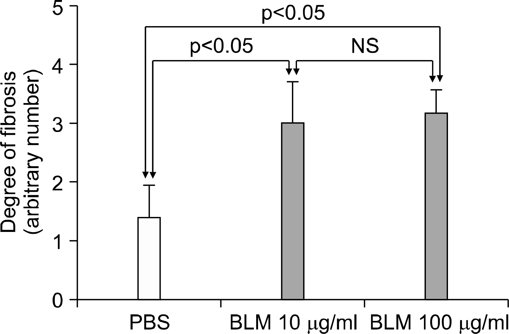
 | Fig. 1.Histopathologic comparison of dermal thickness and fibrosis in C3H/He mice. Mice were treated with PBS 100 μg/ml (1), bleomycin 10 μg/ml (2), or bleomycin 100 μg/ml (3) for 28 days (A: H&E stain, B: Masson's Trichrome stain, magnification ×100). There was no dermal sclerosis or fibrosis in the PBS-treated group. However, bleomycin-treated mice showed increase of dermal thickness and fibrosis. The dermis from the bleomycin-treated group was filled with thickened and homogeneous collagen bundles with cellular infiltrates. |
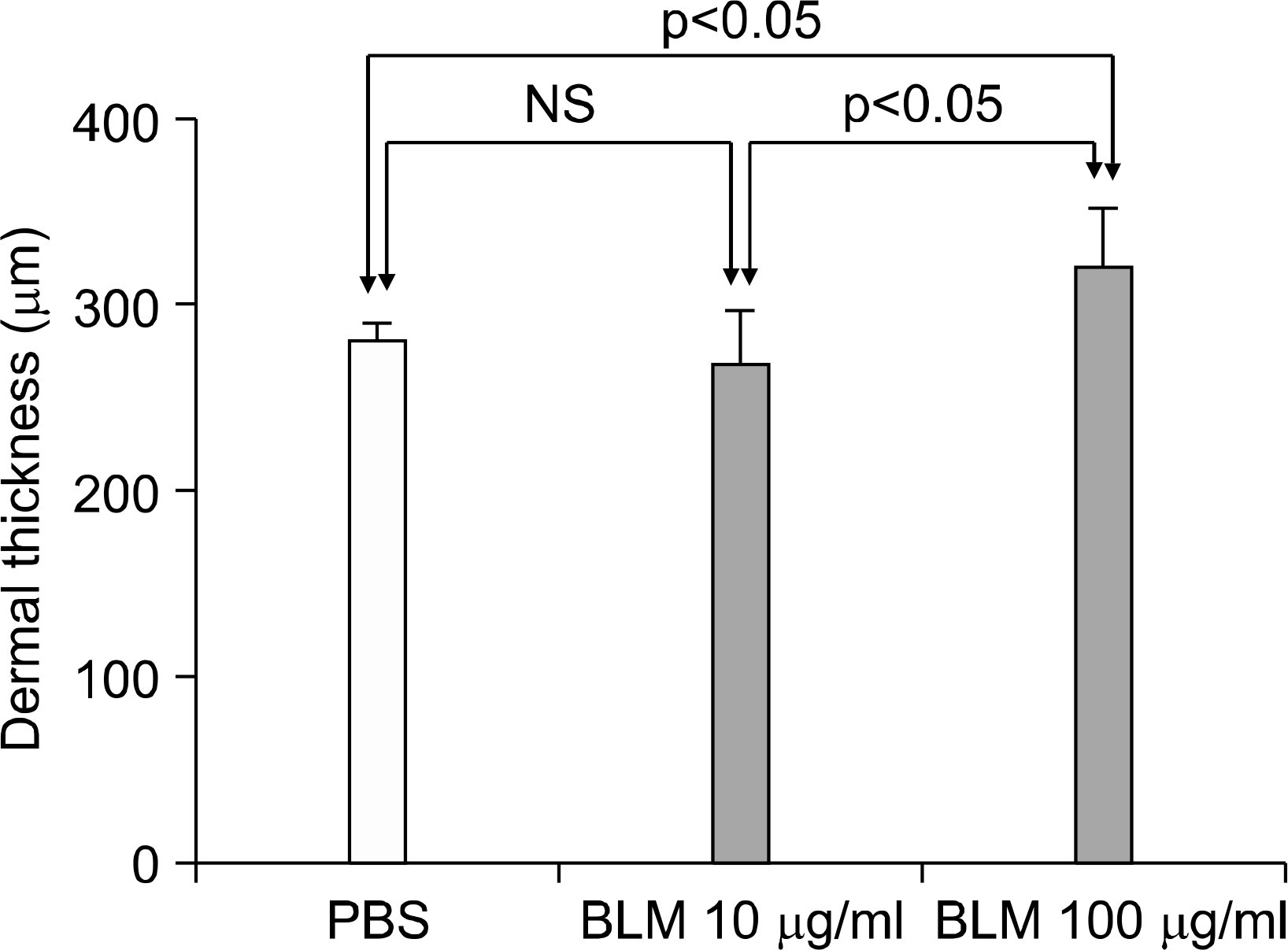 | Fig. 2.Dermal thickness in PBS- or bleomycin-treated mice. H-E stained skin section of mice were evaluated by two pathologists blinded to the treatment group. Repeated injection of bleomycin (10 μg/ml) did not affect dermal thickness; however, 100 μg/ ml of bleomycin significantly increased the dermal thickness (by ANOVA). NS: non-significant, PBS: phosphate buffered saline, BLM: bleomycin. |
 XML Download