

 PDF
PDF ePub
ePub Citation
 Print
Print


References
1. Ehling A, Karrer S, Klebl F, Schaffler A, Muller-Ladner U. Therapeutic management of pyoderma gangrenosum. Arthritis rheum. 2004; 50:3076–84.

2. Park KS, Park W, Song JS, Bae SK, Lee YW, Lee D, et al. A case of pyoderma gangrenosum associated with Behçet's disease. J Korean Rheumatism Assocation. 1999; 6:340–45.
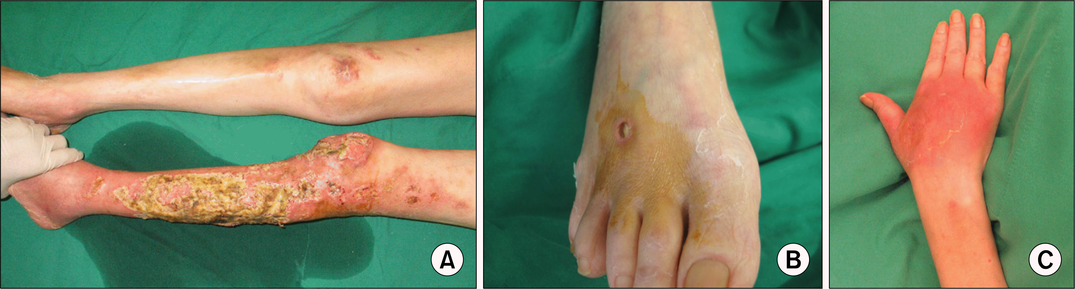
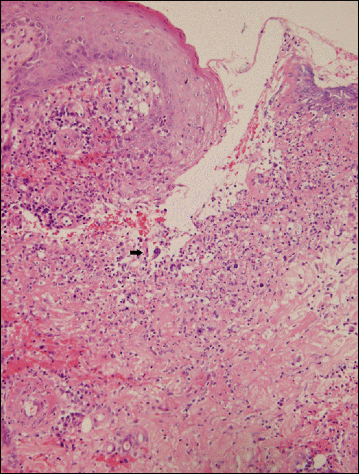
3. Su WP, Davis MD, Weenig RH, Powell FC, Perry HO. Pyoderma gangrenosum: clinicopathologic correlation and proposed diagnostic criteria. Int J Dermatol. 2004; 43:790–800.
4. Weenig RH, Davis MD, Dahl PR, Su WP. Skin ulcers misdiagnosed as pyoderma gangrenosum. N Engl J Med. 2002; 347:1412–8.
5. Callen JP, Jackson JM. Pyoderma gangrenosum: an update. Rheum Dis Clin North Am. 2007; 33:787–802.
 XML Download