

 PDF
PDF ePub
ePub Citation
 Print
Print


Abstract
Churg-Strauss syndrome (CSS), also known as allergic granulomatous angiitis, is a rare disorder characterized by the presence of asthma, eosinophilia and small-to-medium-sized vessels vasculitis. The peripheral nervous system is frequently involved in CSS, but central nervous system (CNS) involvement is rare. Furthermore, transverse myelitis (TM) as a presenting symptom in patients with CSS is extremely rare. We report here on a 60-year-old female who presented peripheral eosinophilia, lung eosinophilic infiltration, lung vasculitis, and TM. She was diagnosed as CSS based on clinical manifestation, pathologic findings, and the use of magnetic resonance imaging (MRI).
Go to : 
References
1. Oh SC, Lee YH, Cho JY, Moon JS, Kim HK, Ji HK, et al. A case of churg-strauss syndrome. J Korean Rheum Assoc. 1996; 3:97–101.
2. Yi JS, Jung HY, Choi SW. A case of acute fulminant neuropathy in patient with allergic granulomatosis and angiitis. J Korean Rheum Assoc. 1998; 5:298–303.
3. Noth I, Strek ME, Leff AR. Churg-Strauss syndrome. Lancet. 2003; 361:587–94.

4. Sinico RA, Bottero P. Churg-Strauss angiitis. Best Pract Res Clin Rheumatol. 2009; 23:355–66.
5. Burns TM, Schaublin GA, Dyck PJ. Vasculitic neuropathies. Neurol Clin. 2007; 25:89–113.
6. Brinar VV, Habek M, Brinar M, MalojcićB , Boban M. The differential diagnosis of acute transverse myelitis. Clin Neurol Neurosurg. 2006; 108:278–83.
7. Oh MJ, Lee JY, Kwon NH, Choi DC. Churg-Strauss syndrome: the clinical features and longterm followup of 17 patients. J Korean Med Sci. 2006; 21:265–71.
8. Kang DH, Jeong IK, Kim HJ, Lee KW. Neurologic Manifestations of Churg-Strauss Syndrome. J Korean Neurol Assoc. 1999; 17:836–40.
9. Seo CG, Kim SH, Lee SH, Yun TS, Park JH, Lim JG. A case of transverse myelitis associated with systemic lupus erythematosus. Keimyung Med J. 2002; 21:100–4.
10. Transverse Myelitis Consortium Working Group. Proposed diagnostic criteria and nosology of acute transverse myelitis. Neurology. 2002; 59:499–505.
11. Sinico RA, Di Toma L, Maggiore U, Bottero P, Radice A, Tosoni C, et al. Prevalence and clinical significance of antineutrophil cytoplasmic antibodies in Churg-Strauss syndrome. Arthritis Rheum. 2005; 52:2926–35.
12. Ozaki S. ANCA-associated vasculitis: diagnostic and therapeutic strategy. Allergol Int. 2007; 56:87–96.
13. Taniguchi M, Tsurikisawa N, Higashi N, Saito H, Mita H, Mori A, et al. Treatment for Churg-Strauss syndrome: induction of remission and efficacy of intravenous immunoglobulin therapy. Allergol Int. 2007; 56:97–103.
14. Lopez Dupla M, Khamashta MA, Sanchez AD, Ingles FP, Uriol PL, Aguado AG. Transverse myelitis as a first manifestation of systemic lupus erythematosus: a case report. Lupus. 1995; 4:239–42.
Go to :
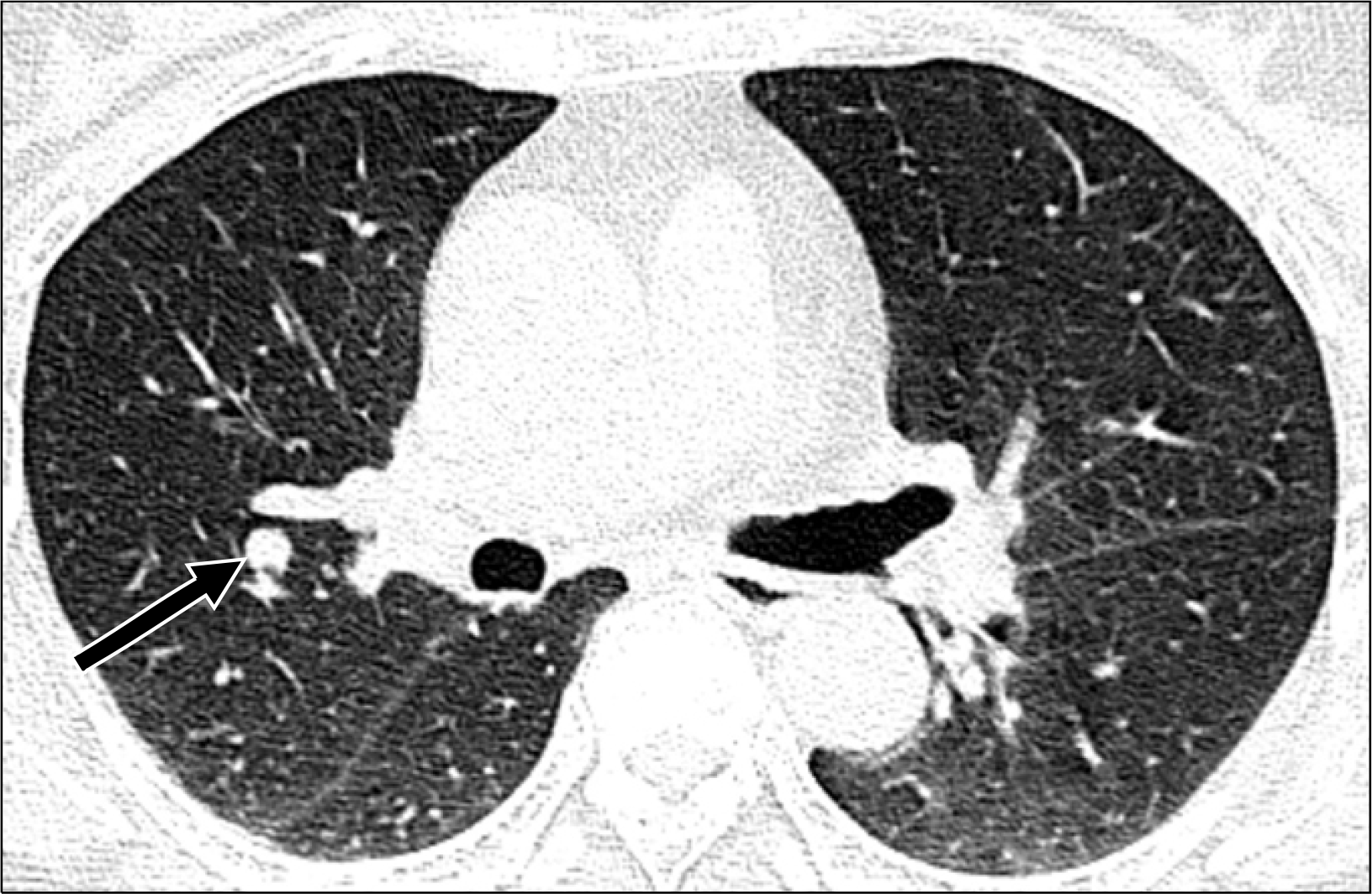 | Fig. 1.Lung setting computed tomography (CT) scan of the chest reveals a well defined nodule in the right upper lobe (0.7 cm in size, arrow). |
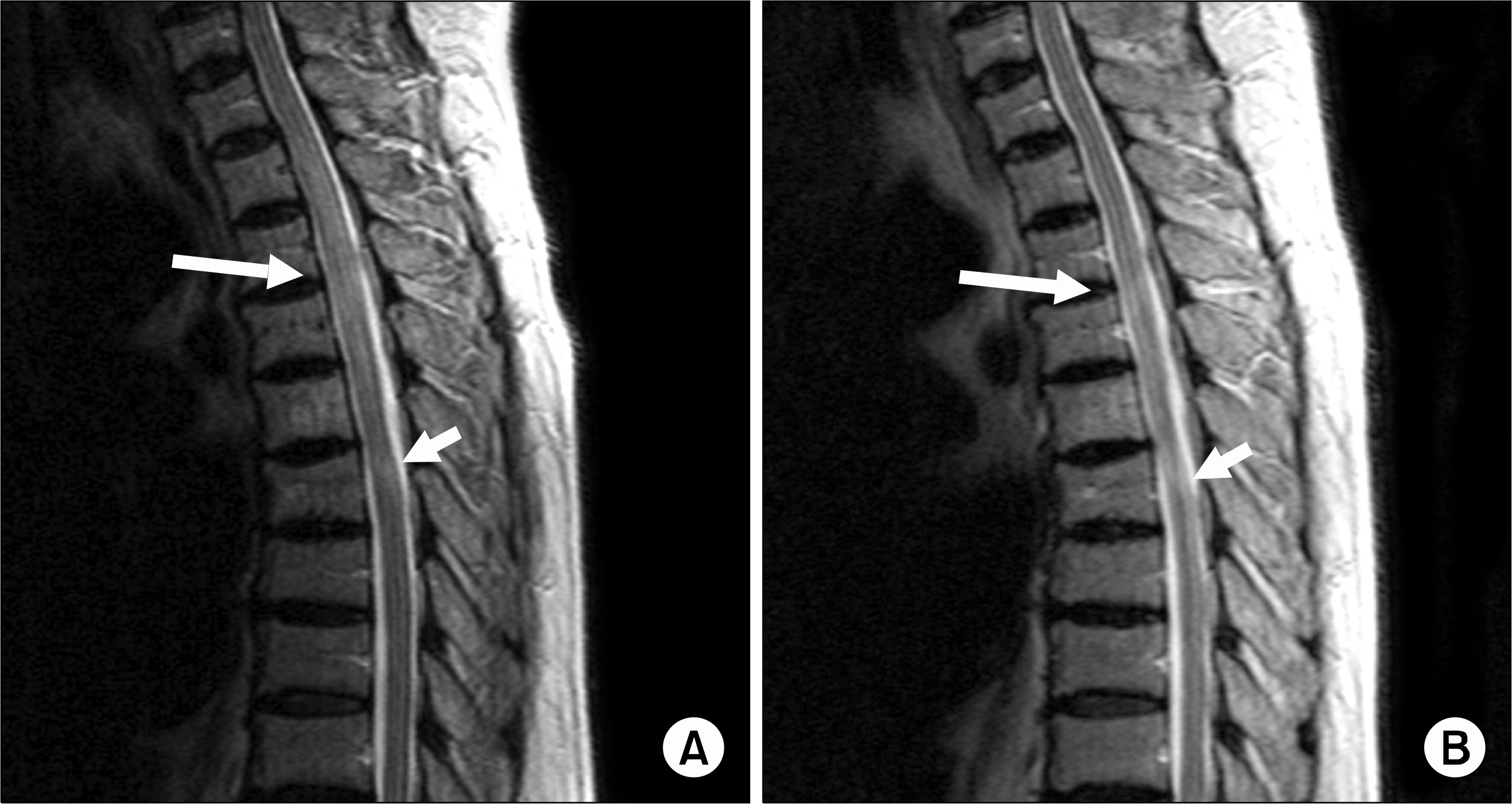 | Fig. 2.MRI findings. (A) Sagittal T2 weighted image (T2WI) of the T spine reveals diffuse swelling and increased signal intensity in T2–5 spinal cord (arrow), and focal increased signal intensity lesion in T6 spinal cord (arrowhead). (B) A four-month follow-up sagittal T2WI of the T spine reveals more resolution in the previous lesions, but there are focal residual signal intensity lesions in T4 and 6 spinal cord (arrow and arrowhead). |

 XML Download