

 PDF
PDF ePub
ePub Citation
 Print
Print


Abstract
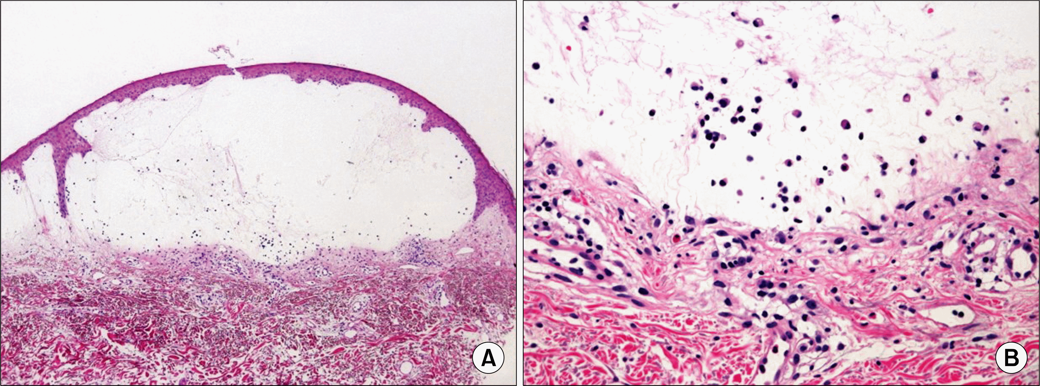
Dermatomyositis is a rare inflammatory myopathy with characteristic skin manifestations and accompanied by muscular weakness. Vesicle formation in dermatomyositis is rare. We report a case of dermatomyositis associated with ovarian cancer in a 62-year-old woman who had vesicles and bullae on her arms. She had erythema and edema on the face, chest, abdomen, and shoulder for 2 months. Diagnosis of dermatomyositis was established by clinical manifestations, muscle enzyme elevation, and a characteristic electromyogram. She was successfully treated with cyclosporin and high doses of steroids.
References
1. Callen JP. Dermatomyositis. Lancet. 2000; 355:53–7.

2. Kubo M, Sato S, Kitahara H, Tsuchida T, Tamaki K. Vesicle formation in dermatomyositis associated with gynecologic malignancies. J Am Acad Dermatol. 1996; 34:391–4.
3. Lee HS, Choi HU, Bae JH, Lee SK. A case of dermatomyositis associated with esophageal cancer showing vesicular and bullous lesions. Korean J Dermatol. 2002; 40:1538–42.
4. Levine SM. Cancer and myositis: new insights into an old association. Curr Opin Rheumatol. 2006; 18:620–4.
5. Zangrilli A, Papoutsaki M, Bianchi L, Teoli M, Chimenti S. Bullous dermatomyositis: a marker of poor prognosis and aggressive internal malignancy? Acta Derm Venereol. 2008; 88:393–4.
6. Smith ES, Hallman JR, DeLuca AM, Goldenberg G, Jorizzo JL, Sangueza OP. Dermatomyositis: a clinicopathological study of 40 patients. Am J Dermatopathol. 2009; 31:61–7.
7. McCollough ML, Cockerell CJ. Vesiculo-bullous dermatomyositis. Am J Dermatopathol. 1998; 20:170–4.

 XML Download