

 PDF
PDF ePub
ePub Citation
 Print
Print


Abstract
Systemic lupus erythematosus (SLE) is a multisystemic inflammatory autoimmune disease caused by various autoantibodies and immune complexes. SLE and antiphospholipid antibodies are associated with thrombotic manifestations. However, renal artery thrombosis which causes renal artery occlusion is uncommon even in SLE patients with antiphospholipid antibodies. A 27-year-old woman with SLE suddenly developed left flank pain and generalized edema. From the laboratory workup, the woman was negative for antiphospholipid antibody and nephrotic-range proteinuria was detected. Computed tomography revealed renal artery thromboembolism and multiple renal infarctions with parenchymal perfusion defects in the left kidney. Renal biopsy showed WHO classification III and V lupus nephritis. Left flank pain, generalized edema and proteinuria were resolved and the thromboembolism resolved itself after a high dose of steroid and anticoagulation therapy. In SLE patients, sudden onset of unexplained flank pain is considered as a possible symptom of renal vessel thromboembolism even if the antiphospholipid antibody is negative.
References
1. Mok CC, Tang SS, To CH, Petri M. Incidence and risk factors of thromboembolism in systemic lupus erythematosus: a comparison of three ethnic groups. Arthritis Rheum. 2005; 52:2774–82.

2. Appel GB, Pirani CL, D'Agati V. Renal vascular complications of systemic lupus erythematosus. J Am Soc Nephrol. 1994; 4:1499–515.
3. Frostegard J. SLE, atherosclerosis and cardiovascular disease. J Intern Med. 2005; 257:485–95.
4. Tsugawa K, Tanaka H, Kudo M, Nakahata T. Renal artery thrombosis in a pediatric case of systemic lupus erythematosus without antiphospholipid antibodies. Pediatr Nephrol. 2005; 20:1648–50.
5. Suwabe H, Moriuchi J, Hoshina Y. Renal and cerebral infarctions in a patient with systemic lupus erythematosus without antiphospholipid antibodies. Ryumachi. 1993; 33:335–40.
6. Hall S, Buettner H, Luthra HS. Occlusive retinal vascular disease in systemic lupus erythematosus. J Rheumatol. 1984; 11:846–50.
7. Nikpour M, Urowitz MB, Gladman DD. Premature atherosclerosis in systemic lupus erythematosus. Rheum Dis Clin North Am. 2005; 31:329–54.
8. Asherson RA, Derksen RH, Harris EN, Bouma BN, Gharavi AE, Kater L, et al. Chorea in systemic lupus erythematosus and "lupus-like" disease: association with antiphospholipid antibodies. Semin Arthritis Rheum. 1987; 16:253–9.
9. Hernandez D, Dominquez ML, Diaz F, Fernandez ML, Lorenzo V, Rodriquez A, et al. Renal infarction in a severely hypertensive patient with lupus erythematosus and antiphospholipid antibodies. Nephron. 1996; 72:298–301.
10. Laskin CA, Clark CA, Spitzer KA. Antiphospholipid syndrome in systemic lupus erythematosus: is the whole greater than the sum of its parts? Rheum Dis Clin North Am. 2005; 31:255–72.
11. Soltesz P, Veres K, Lakos G, Kiss E, Muszbek L, Szeqedi G. Evaluation of clinical and laboratory features of antiphospholipid syndrome: a retrospective study of 637 patients. Lupus. 2003; 12:302–7.
12. Raij L, Shultz PJ. Endothelium-derived relaxing factor, nitric oxide: effects on and production by mesangial cells and the glomerulus. J Am Soc Nephrol. 1993; 3:1435–41.
13. Ross R. Atherosclerosis–an inflammatory disease. N Engl J Med. 1999; 340:115–26.
Fig. 1.
27-year-old woman with multiple left renal infarctions. (A) Contrast-enhanced computed tomography image during corticomedullary phase shows multiple parenchymal perfusion defects in the left kidney compatible with renal infarctions. Multiple filling defects (arrows) in left main and segmental renal arteries suggest the thromboembolism. (B) Follow-up contrast-enhanced maximal intensity projection image obtained one week later shows the thromboembolism is no longer present in the left main and segmental renal arteries as well as a decreased extent of parenchymal loss.
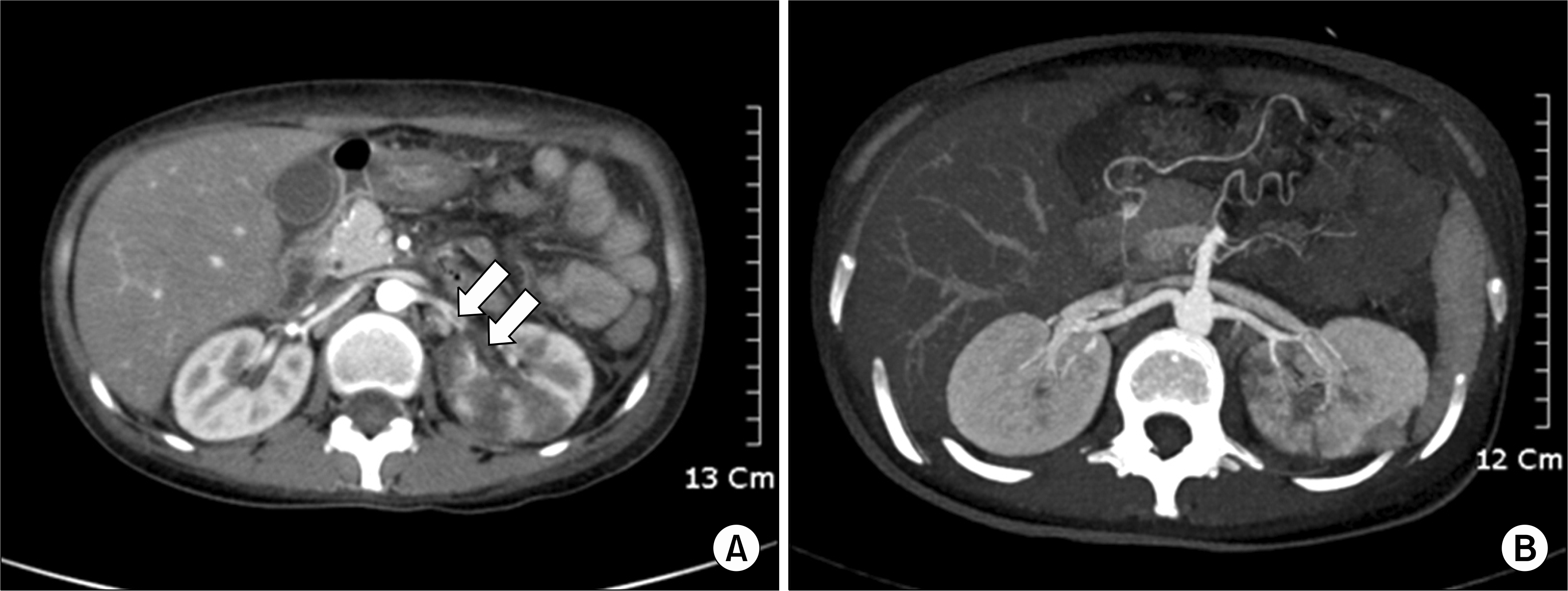
Fig. 2.
27-year-old woman with lupus nephritis WHO class III+V. (A) Light microscopy shows moderate hypercellular mesangial tissue and focal endocapillary proliferation. Capillary loops are focally thickened with subendothelial deposition (H&E stain, ×400). (B) Electron microscopy shows diffuse effacement of epithelial foot processes and wide spread subepithelial electron-dense deposition (×5,000).
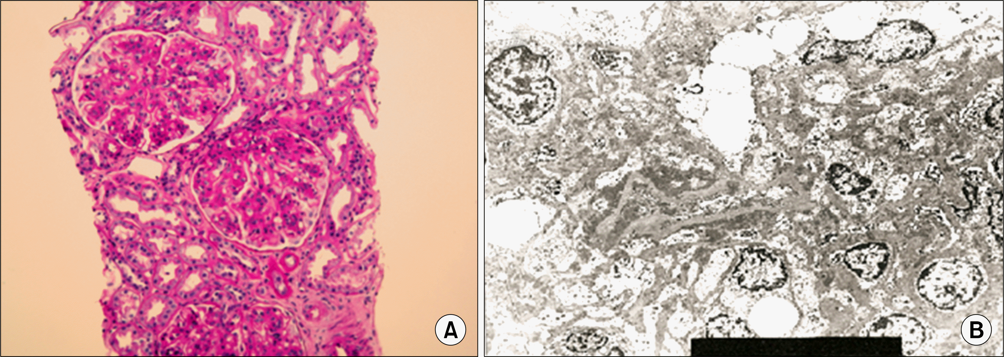
Table 1.
Comparison of previous renal infarction cases of SLE patients negative for antiphospholipid antibodies to the current case
| A∗ | B† | C‡ | |
|---|---|---|---|
| Sex | Male | Female | Female |
| Race | Asian | Asian | Asian |
| Age | 13-year old | 42-year-old | 27-year-old |
| SLE duration | 11-month | 8-year | 4-year |
| Symptom | Hypertension, fever, seizure, abdominal pain | Right abdominal pain | Left flank pain, generalized edema |
| Drug history | Prednisolone, mizoribine, dilazep dihydrochloride | Prednisolone, azathioprine | Prednisolone, hydroxychloroquine |
| Other complication | CNS lupus, lupus nephritis | Lupus nephritis | Lupus nephritis |
∗ Tsugawa K, Tanaka H, Kudo M, Nakahata T. Renal artery thrombosis in a pediatric case of systemic lupus
 XML Download