Abstract
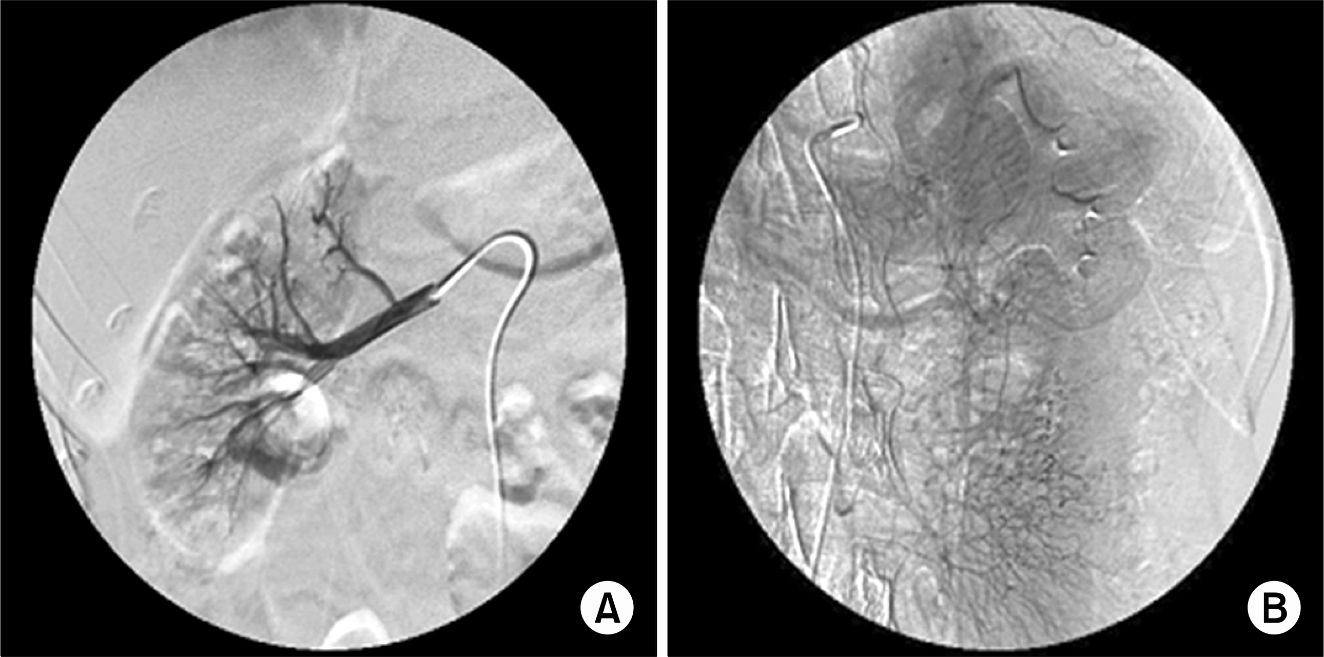
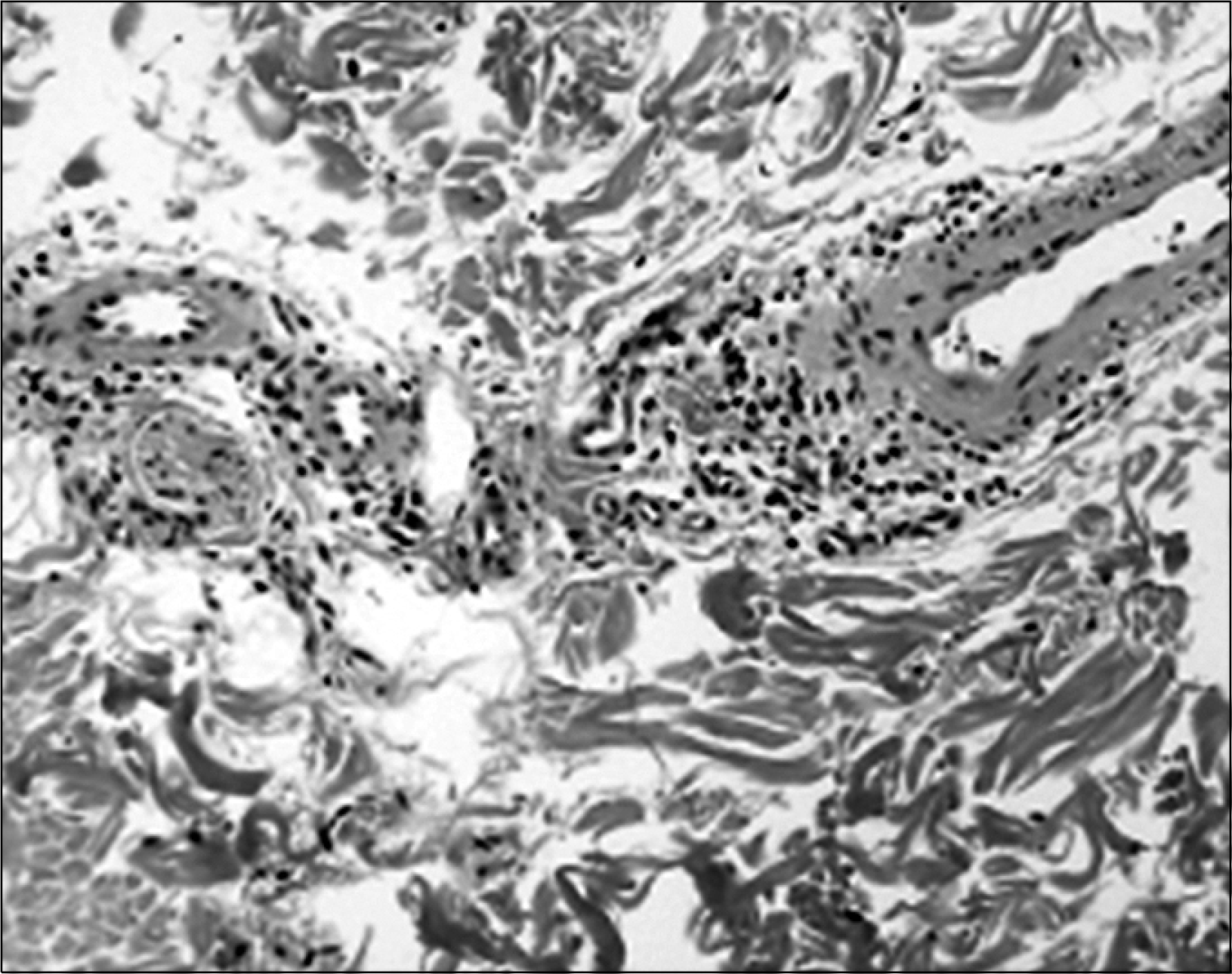
We describe a 28-year old man in otherwise apparently good health, in whom pain in his left knee joint caused by avascular necrosis led to a diagnosis of polyarteritis nodosa (PAN). The angiogram showed multiple microaneurysmal and thrombotic lesions, notably in the renal, mesenteric and tibial arteries. A skin biopsy of the upper dermis of the left thigh with an erythematous skin rash showed the infiltration of mononuclear leukocytes in the perivascular area. During hospitalization, he was diagnosed with chronic hepatitis B, and was treated with lamivudine, and corticosteroid, azathioprine to control the PAN. The knee joint pain improved progressively, and the patient could walk normally after several months. This case is an unusual presentation because the initial manifestation of PAN was avascular necrosis.
Go to : 
References
1. Dubois EL, Cozen L. Avascular bone necrosis associated with systemic lupus erythematosus. JAMA. 1960; 174:966–71.
2. Assouline-Dayan Y, Chang C, Greenspan A, Shoenfeld Y, Gershwin ME. Pathogenesis and natural history of osteonecrosis. Semin Arthritis Rheum. 2002; 32:94–124.

3. Abeles M, Urman JD, Rothfield NF. Aseptic necrosis of bone in systemic lupus erythematosus. Relationship to corticosteroid therapy. Arch Intern Med. 1978; 138:750–4.

4. Sergent JS, Lockshin MD, Christian CL, Gocke DJ. Vasculitis with hepatitis B antigenemia: longterm observation in nine patients. Medicine (Baltimore). 1976; 55:1–18.
5. Mont MA, Glueck CJ, Pacheco IH, Wang P, Hungerford DS, Petri M. Risk factors for osteonecrosis in systemic lupus erythematous. J Rheumatol. 1997; 24:654–62.
6. Gladman DD, Urowits MB, Chaudhry-Ahluwalia V, Hallet DC, Cook RJ. Predictive factors for symptomatic osteonecrosis in patients with systemic lupus erythematous. J Rheumatol. 2001; 28:761–5.
7. Mok CC, Lau CS, Wong RWS. Risk factors for avascular bone necrosis in systemic lupus erythematosus. Br J Rheumatol. 1998; 37:895–900.

8. Heron E, Fiessinger JN, Guillevin L. Polyarteritis nodosa presenting as acute leg ischemia. J Rheumatol. 2003; 30:1344–6.
9. Hall C, Mongey AB. Unusual presentation of polyarteritis nodosa. J Rheumatol. 2001; 28:871–3.
10. Choi SW, Lew S, Cho SD, Cha HJ, Eum EA, Jung HC, et al. Cutaneous polyarteritis nodosa presented with digital gangrene: a case report. J Korean Mes Sci. 2006; 21:371–3.

11. Cohen RD, Conn DL, Ilstrup DM. Clinical features, prognosis and response to treatment in polyarteritis. Mayo Clin Proc. 1980; 55:146–55.
12. Leib ES, Restivo C, Paulus HE. Immunosuppressive and corticosteroid therapy of polyarteritis nodosa. Am J Med. 1979; 67:941–7.

13. Guillevin L, Mahr A, Callard P, Godmer P, Pagnoux C, Leray E, et al. Hepatitis B virus-associated polyarteritis nodosa clinical characteristics, outcome, and impact of treatment in 115 patients. Medicine (Baltimore). 2005; 84:313–22.
Go to : 
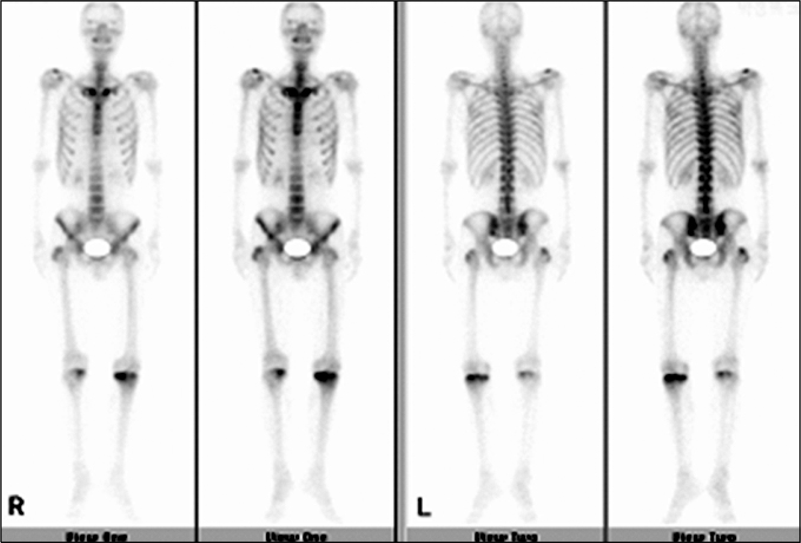
 | Fig. 1.Whole body bone scan showing increased uptake lesion in the left and right upper tibia, indicating osteonecrotic changes. |
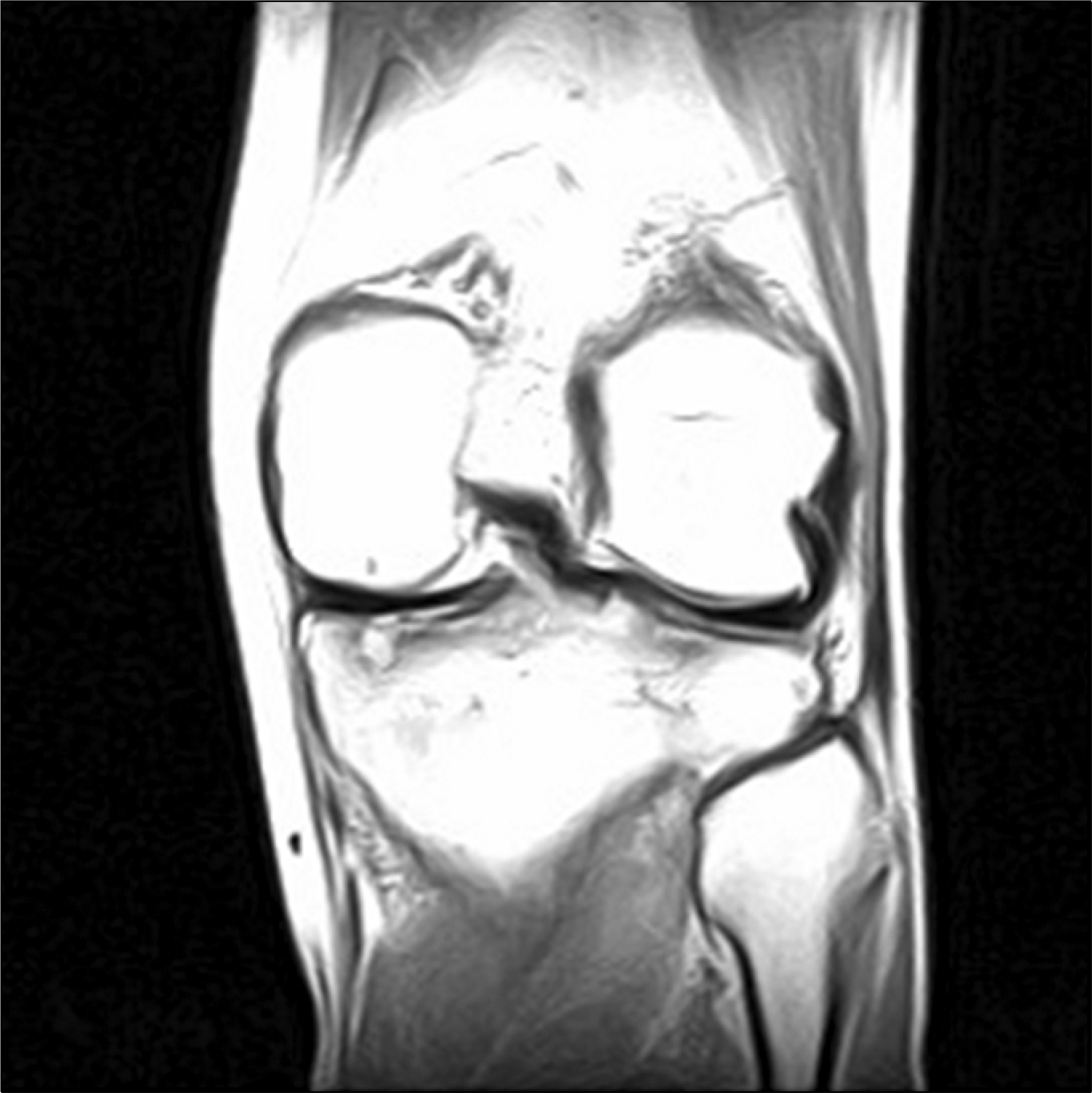
 | Fig. 2.Magnetic resonance imaging of the left knee. The coronal view of the proton image shows an irregular articular margin with bone cortical necrotic changes and diffuse low signal intensity in the upper tibia. |


 PDF
PDF ePub
ePub Citation
 Print
Print


 XML Download