Abstract
Objective
Mature B cells in the spleen of mouse can be divide into two main subsets: the follicular (FO) B cells and the marginal zone (MZ) B cells. In this study, we investigated which subtype of B cells is involved in the production of costimulatory molecules, cytokine and antibody during the induction of autoimmune arthritis.
Methods
The MZB and FOB cells isolated from DBA/1J induced- and collagen-induced arthritis (CIA) mice were stimulated with LPS or CpG. The costimulatory molecules were measured by flow cytometry (FACs). The cytokines were measured by ELISA. Production of antibodies by the MZB cells or FOB cells was measured by ELISA and the results were observed by confocal microscopy.
Results
The expression of co-stimulatory molecules was stronger in the MZB cells than that in the FOB cells. The production of cytokines (IL-10, IL-6) and antibodies was higher in the MZB cells. The IgG expression of the MZB cells, which is known to be associated with the acceleration of autoimmunity, was higher in the CIA mice than that in the DBA/1J mice.
Conclusion
We observed that the MZB cells were increased in the CIA mice. The costimulatory molecules, cytokine and auto-antibodies were increased in the MZB cells compared to that of the FOB cells. Our results suggest that MZB cells mainly produce autoantibodies, and they play a key role in development of autoimmune arthritis.
Go to : 
References
1. Wang Y, Zhang P, Li W, Hou L, Wang J, Liang Y, et al. Mouse follicular and marginal zone B cells show differential expression of Dnmt3a and sensitivity to 5'-azacytidine. Immunol Lett. 2006; 105:174–9.

2. Oliver AM, Martin F, Gartland GL, Carter RH, Kearney JF. Marginal zone B cells exhibit unique activation, proliferative and immunoglobulin secretory responses. Eur J Immunol. 1997; 27:2366–74.

3. Martin F, Kearney JF. B-cell subsets and the mature preimmune repertoire. Marginal zone and B1 B cells as part of a "natural immune memory". Immunol Rev. 2000; 175:70–9.
7. Gunn KE, Brewer JW. Evidence that marginal zone B cells possess an enhanced secretory apparatus and exhibit superior secretory activity. J Immunol. 2006; 177:3791–8.

8. Cinamon G, Zachariah MA, Lam OM, Foss FW Jr, Cyster JG. Follicular shuttling of marginal zone B cells facilitates antigen transport. Nat Immunol. 2008; 9:54–62.

9. Oliver AM, Martin F, Kearney JF. IgMhighCD21high lymphocytes enriched in the splenic marginal zone generate effector cells more rapidly than the bulk of follicular B cells. J Immunol. 1999; 162:7198–207.
10. Grimaldi CM, Michael DJ, Diamond B. Cutting edge: expansion and activation of a population of autoreactive marginal zone B cells in a model of estrogen-induced lupus. J Immunol. 2001; 167:1886–90.

11. Barnett ML, Kremer JM, St Clair EW, Clegg DO, Furst D, Weisman M, et al. Treatment of rheumatoid arthritis with oral type II collagen. Results of a multicenter, double-blind, placebocontrolled trial. Arthritis Rheum. 1998; 41:290–7.

Go to : 
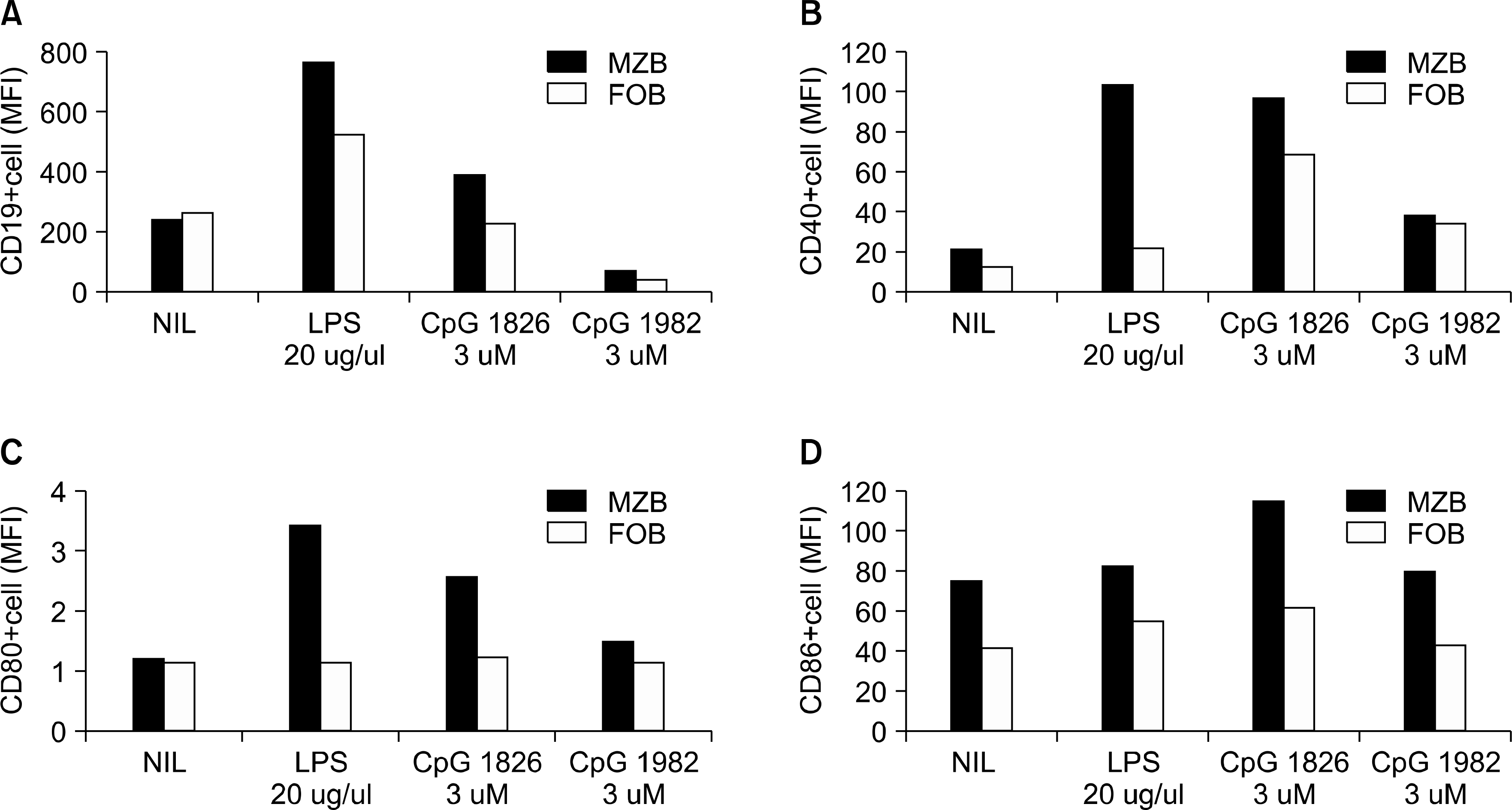 | Fig. 1.The expressions of the CD19 and costimulatory molecules (CD40, CD80, CD86) in the MZB cells and FOB cells. The freshly isolated MZB cells (black bar) and FOB cells (white bar) that were cultured for 24 hours in vitro from the DBA/1J mice were analyzed by flowcytometry. (A) CD19, (B) CD40, (C) CD80 and (D) CD86. |
 | Fig. 2.Evaluation of the cytokine production in the culture supernatants from the MZB and FOB cells. Freshly isolated MZB (black bar) and FOB cells (white bar) from DBA/1J mice wete cultured for 24 hours in vitro. The IL-10 (A) and IL-6 (B) production was measured by ELISA. The Values are the mean±standard deviation from three independent experiments. ∗p<0.01. |
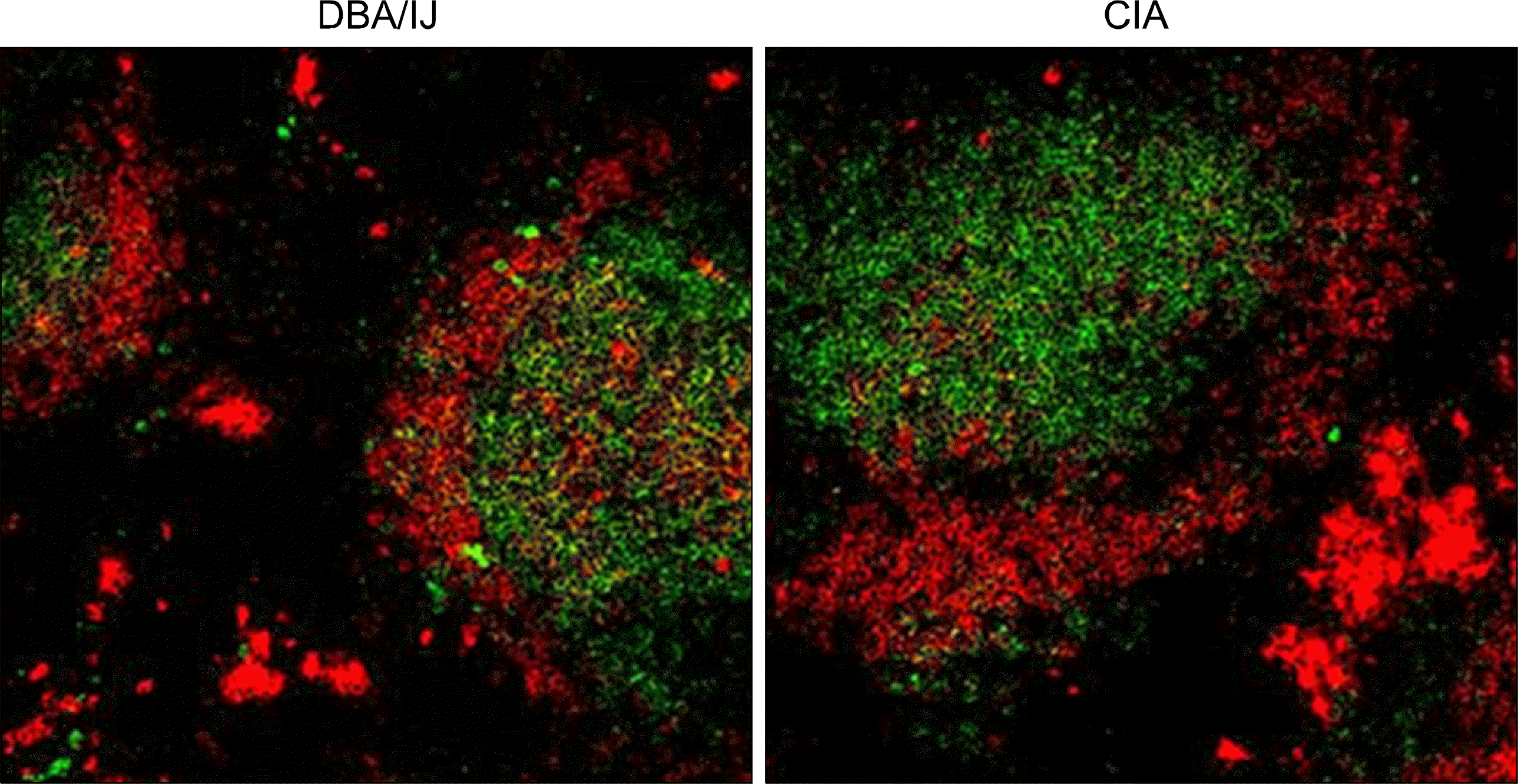 | Fig. 3.Confocal microscopy analysis of the MZB cells and FOB cells in the DBA/1J and CIA mice spleens. Cryo-sections of the spleens from the DBA/1J mice and CIA mice were stained by anti-mouse IgD (FITC conjugated, green), anti-mouse IgM (Biotin) and streptavidin-Cy3 (red) antibodies. We analyzed this on confocal microscopy (×200). |
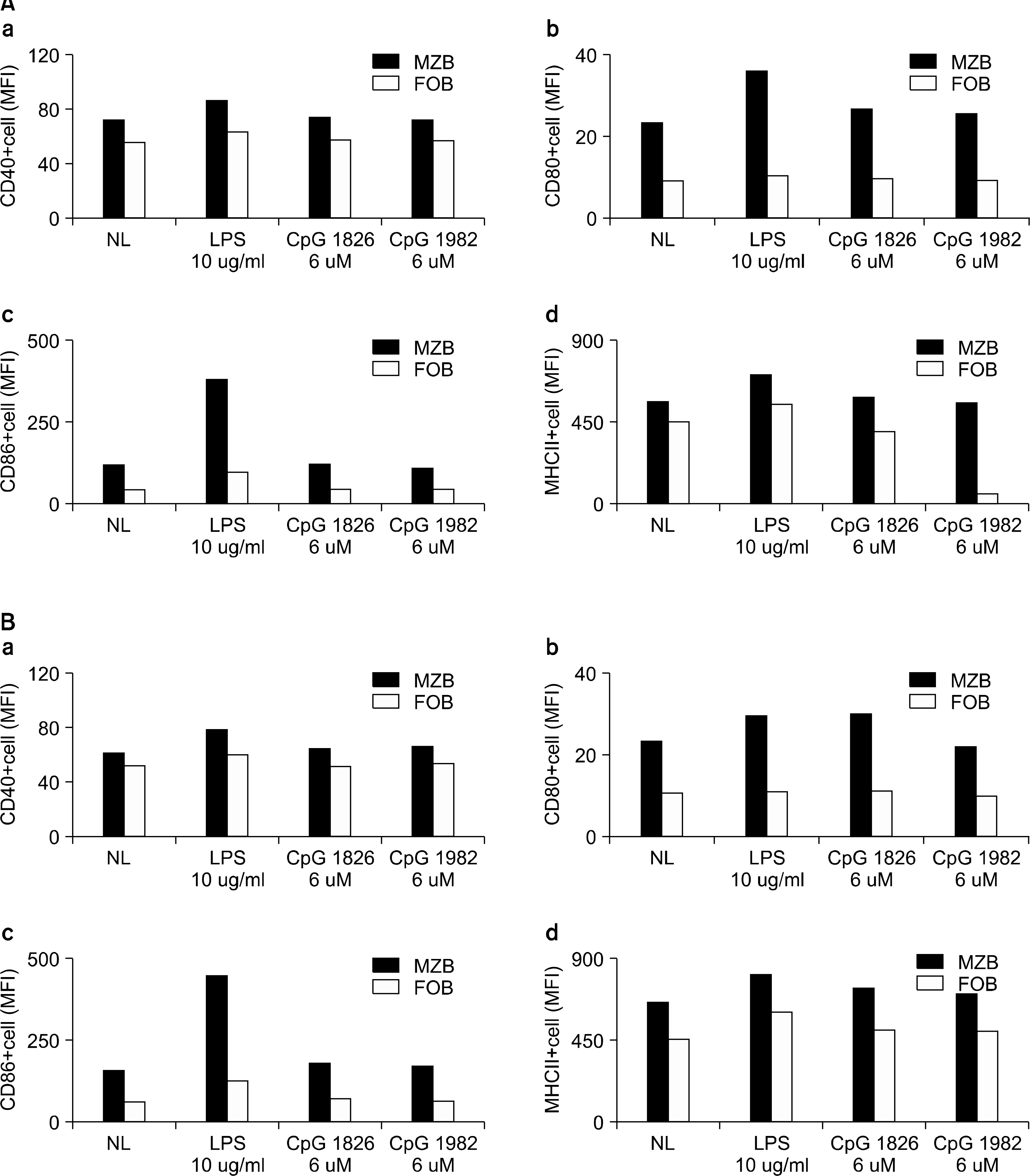 | Fig. 4.The expression of costimulatory molecules (CD40, CD80, CD86, MHCII) in the MZB and FOB cells from the DBA/1J and CIA mice. Freshly isolated splenocytes from the DBA/1J mice (A) and CIA mice (B) were cultured with LPS, CpG1826 or CpG1982 for 6 hours. The proportions of MZB cells and FOB cells were determined using FACs. (a) CD40, (b) CD80, (c) CD86 and (d) MHCII. |
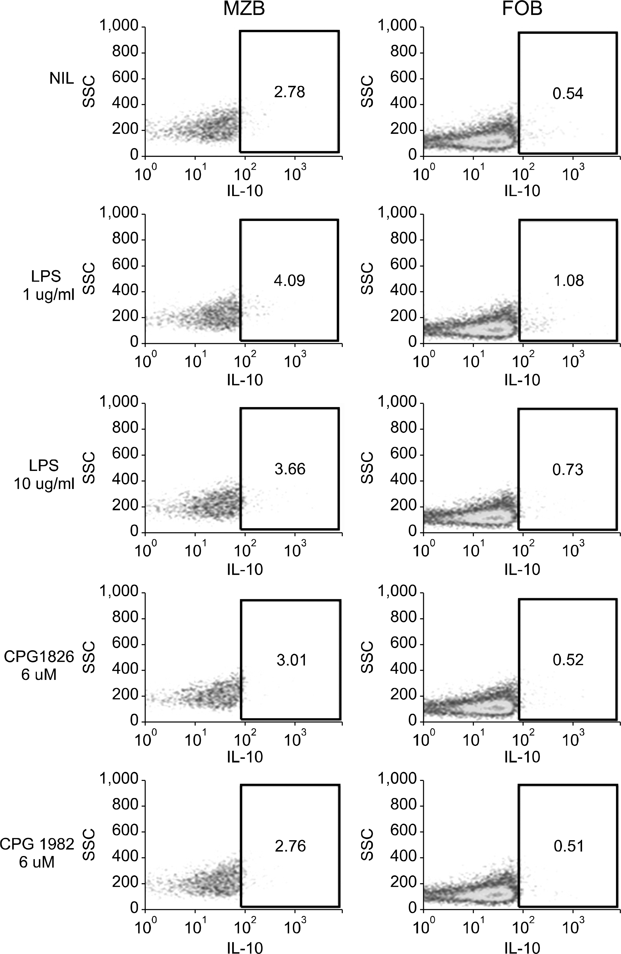 | Fig. 5.FACs analysis of IL-10 in MZB and FOB cells from CIA mice. Freshly isolated MZB and FOB cells cultured for 6 hours in vitro from CIA mice. IL-10 production was measured by flowcytometry. |
 | Fig. 6.IgG and IgM production was measured in culture supernatants from MZB and FOB cells. Freshly isolated MZB (black bar) and FOB cells (white bar) cultured for 24 hours in vitro from DBA/1J mice. IgM (A) and IgG (B) production were measured by ELISA. Values are the mean±standard deviation from three independent experiments. ∗p<0.01. |
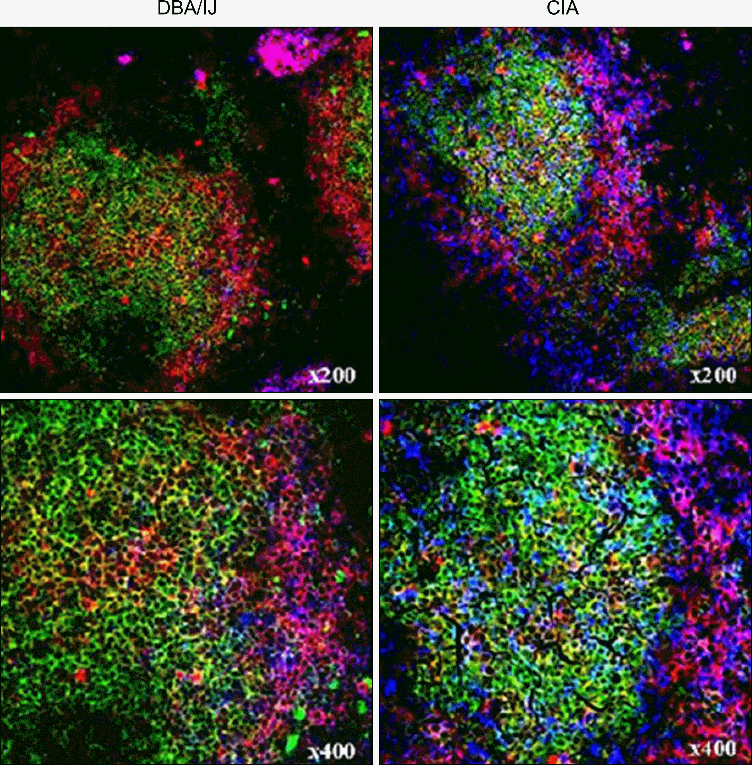 | Fig. 7.The IgG expression of the MZB cells and FOB cells in the DBA/1J and CIA mice spleens. Cryo-sections of the spleen from the DBA/1J mice and CIA mice were stained by anti-mouse IgD (FITC conjugated, green), anti-mouse IgM (Biotin), streptavidin-Cy3 (red) and anti-mouse IgG (APC conjugated, blue) antibodies. We analyzed the results on confocal microscopy. The merged IgM+ IgG+ cells were increased in the CIA mice. |


 PDF
PDF ePub
ePub Citation
 Print
Print


 XML Download