Abstract
Reduced bone mineral density precedes the development of vertebral fractures in patients under long term glucocorticoid therapy. Osteoporosis is a frequent complication in steroid-dependent patients, and the risk of developing vertebral fractures in these patients is much higher than involutional osteoporosis. We described a 54-year-old patient who presented with autoimmune hepatitis and had a 6-year history of steroid medication. The patient had multiple compression fractures (T10∼L5) without trauma, and was treated successfully with multi-level vertebroplasty and an intravenous injection of bisphosphonate without complications.
References
1. Stellon AJ, Webb A, Compston JE. Bone histomorphometry and structure in corticosteroid treated chronic active hepatitis. Gut. 1988; 29:378–84.

2. Jehle PM. Steroid induced osteoporosis: how can it be avoided? Nephrol Dial Transplant. 2003; 18:861–4.
3. Reid IR. Time to end steroid induced fractures. Arch Dermatol. 2001; 137:493–4.
4. Choi WH, Kim SY, Kim TH. Vertebral fracture in long term steroid dependent patients. 1994; 1:224–9.
5. Lim SH, Kim H, Kim HK, Baek MJ. Multiple cardiac perforations and pulmonary embolism caused by cement leakage after percutaneous vetebroplasty. Eur J Cardiothirac Surg. 2008; 33:510–2.
6. Kim YJ, Lee JW, Park KW, Yeom JS, Park JM, Kang HS. Pulmonary cement embolism after percutaneous vertebroplasty in osteoporotic vertebral compression fractures: incidence, charicteristics, and risk factors. Radiology. 2009; 251:250–9.
7. Karaktoprak O, Camurdan K, Ozturk C, Ganiyusufoglu K, Aydogan M, Hamzaoglu A. Multiple-level vertebroplasty in patients with vertebral compression fractures from osteodystrophy in chronic liver disease. Acta Orthop Belg. 2008; 74:566–8.
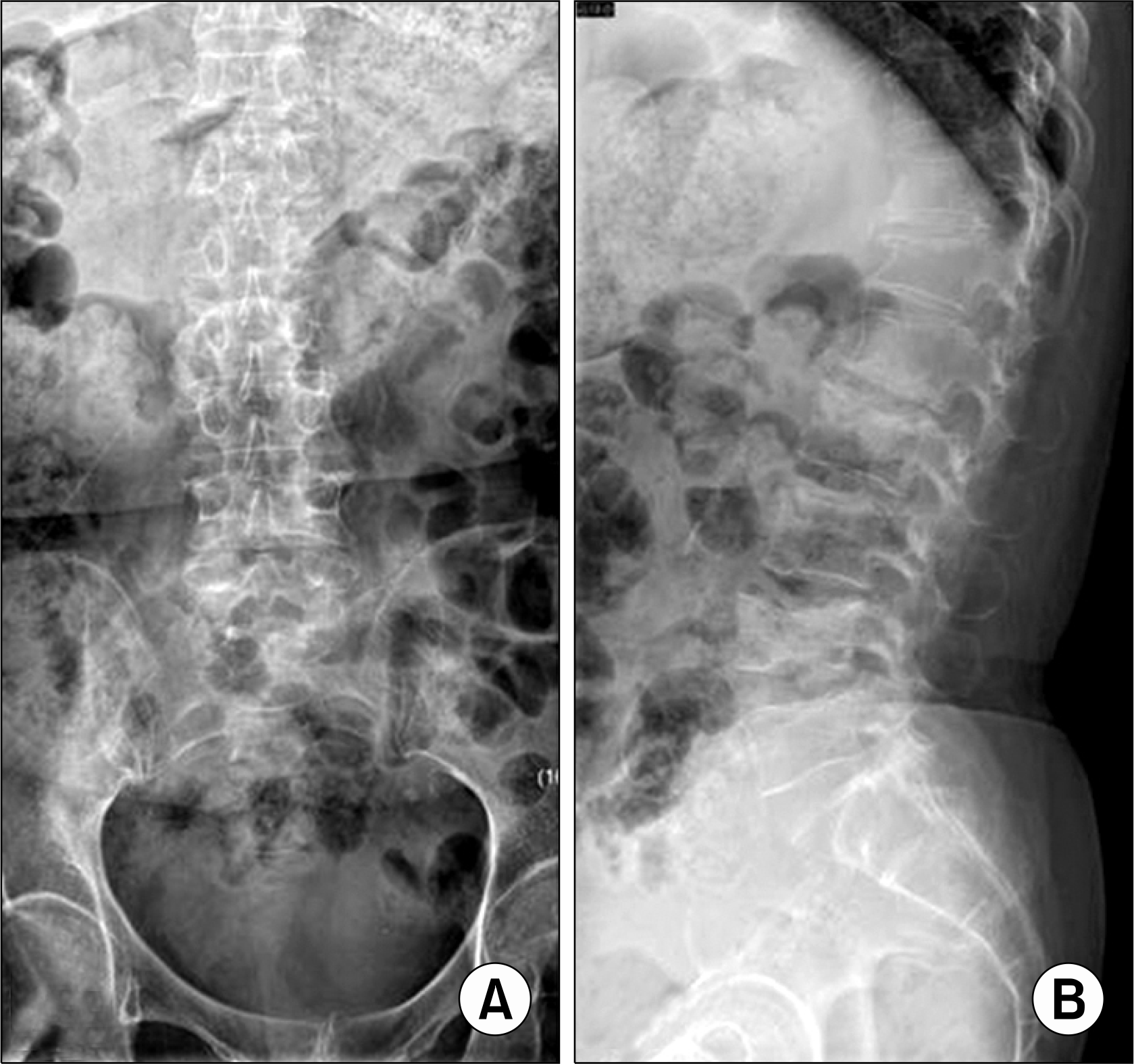
Fig. 1.
Lumbar plain radiography (Pre-operative). Lumbar AP (A) and Lateral (B) x-ray shows multiple compression fractures (T12, L1, 2, 4, 5).

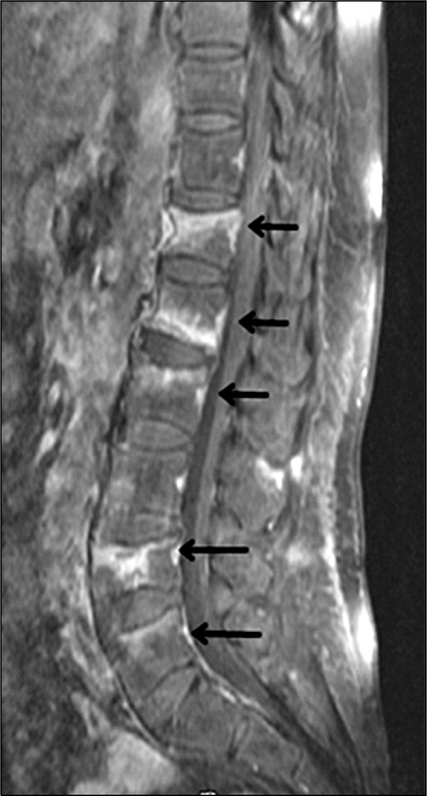
Fig. 2.
Lumbar MRI. Gadolinium-enhanced sagittal image of the lumbar spine shows signal intensity (arrow) of the fractured vertebral segments (T12, L1, 2, 4, 5).


 PDF
PDF ePub
ePub Citation
 Print
Print


 XML Download