Abstract
Cytomegalovirus (CMV) infection commonly affects patients who are in an immunocompromised state, such as acquired immune deficiency syndrome (AIDS) and during organ transplantation. Although cytomegalovirus infection does not occur frequently, it is a major cause of morbidity and mortality in patients suffering with connective tissue diseases, including dermatomyositis. Cytomegalovirus pneumonitis and retinitis has been rarely reported in patients with dermatomyositis. We report here on an usual case involving the simultaneous occurrence of cytomegalovirus pneumonitis and retinitis in a 39-year-old female with dermatomyositis, and this woman had been treated with steroids and immunosuppressive agents for the previous 5 months.
Go to : 
References
1. Kanno M, Chandrasekar PH, Bentley G, Vander Heide RS, Alangaden GJ. Disseminated cytomegalovirus disease in hosts without acquired immunodeficiency syndrome and without an organ transplant. Clin Infect Dis. 2001; 32:313–6.

2. Juárez M, Misischia R, Alarcón GS. Infections in systemic connective tissue diseases: systemic lupus erythematosus, scleroderma, and polymyositis/derma-tomyositis. Rheum Dis Clin North Am. 2003; 29:163–84.

3. Kasifoglu T, Korkmaz C, Ozkan R. Cytomegalovirus-induced interstitial pneumonitis in a patient with dermatomyositis. Clin Rheumatol. 2006; 25:731–3.

4. Kim HR, Kim SD, Kim SH, Yoon CH, Lee SH, Park SH, et al. Cytomegalovirus retinitis in a patient with dermatomyositis. Clin Rheumatol. 2007; 26:801–3.

5. Yoda Y, Hanaoka R, Ide H, Isozaki T, Matsunawa M, Yajima N, et al. Clinical evaluation of patients with inflammatory connective tissue diseases complicated by cytomegalovirus antigenemia. Mod Rheumatol. 2006; 16:137–42.

6. Takizawa Y, Inokuma S, Tanaka Y, Saito K, Atsumi T, Hirakata M, et al. Clinical characteristics of cytomegalovirus infection in rheumatic diseases: multi-centre survey in a large patient population. Rheumatology (Oxford). 2008; 47:1373–8.

7. Reed JB, Schwab IR, Gordon J, Morse LS. Regression of cytomegalovirus retinitis associated with protease-inhibitor treatment in patients with AIDS. Am J Ophthalmol. 1997; 124:199–205.

8. Holland GN, Tufail A, Jordan MC. Cytomegalovirus Diseases. In: Ocular Infection and Immunity. p.1088, St. Louis, CV Mosby. 1996.
9. Goldberg DE, Smithen LM, Angelilli A, Freeman WR. HIV-associated retinopathy in the HAART era. Retina. 2005; 25:633–49.

10. Meyers JD, Flournoy N, Thomas ED. Risk factors for cytomegalovirus infection after human marrow transplantation. J Infect Dis. 1986; 153:478–88.

11. Najjar M, Siddiqui AK, Rossoff L, Cohen RI. Cavitary lung masses in SLE patients: an unusual manifestation of CMV infection. Eur Respir J. 2004; 24:182–4.

12. Karakelides H, Aubry MC, Ryu JH. Cytomegalovirus pneumonia mimicking lung cancer in an immunocompetent host. Mayo Clin Proc. 2003; 78:488–90.

13. Salomon N, Gomez T, Perlman DC, Laya L, Eber C, Mildvan D. Clinical features and outcomes of HIV-related cytomegalovirus pneumonia. AIDS. 1997; 11:319–24.
Go to : 
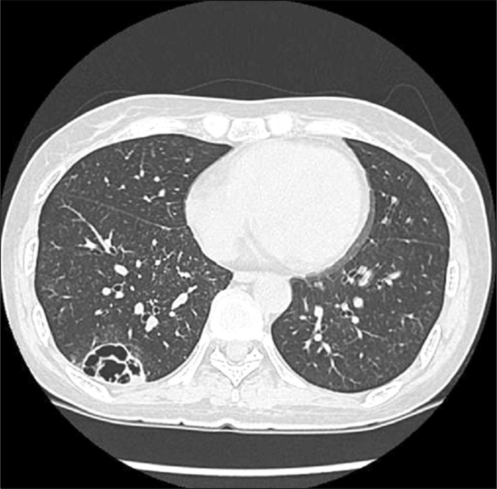
 | Fig. 1.Chest CT shows a cavitary lesion (4.1×4.0 cm) with a thin wall and internal septation at the subpleural region of the right lower lobe. |
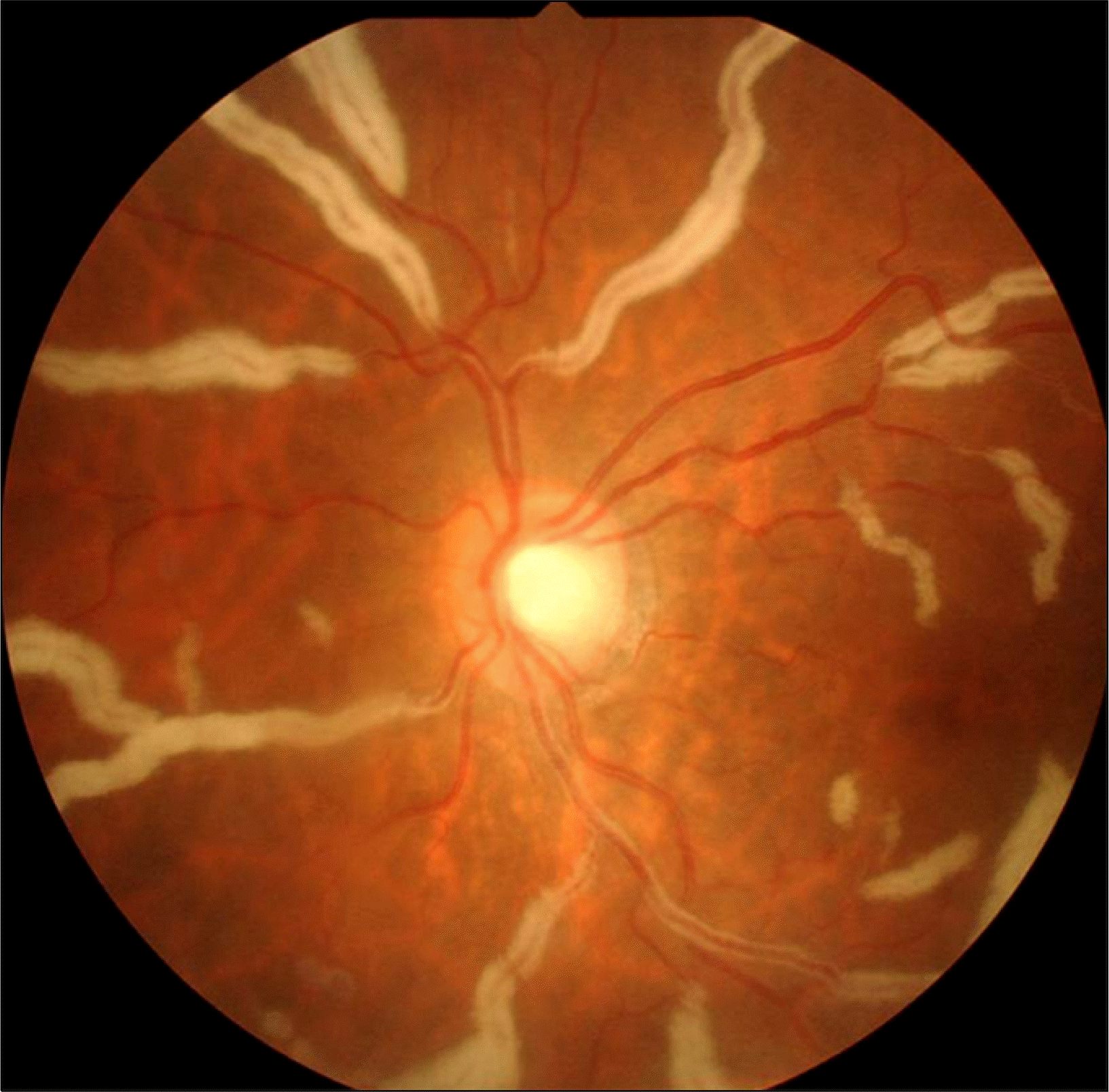
 | Fig. 2.The fundoscopic examination shows retinal whitening and vascular sheathings, and these findings are consistent with CMV retinitis. |
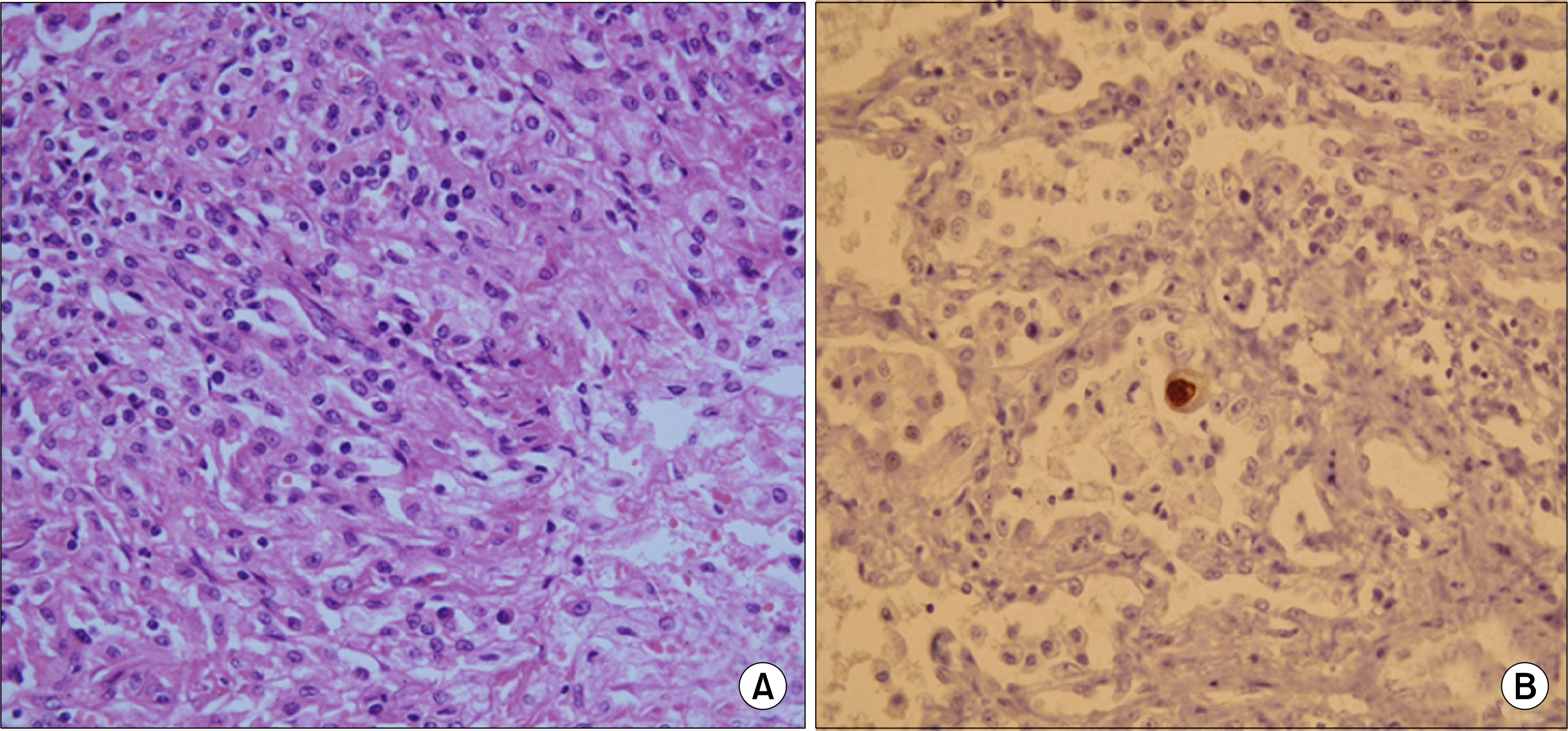
 | Fig. 3.The thoracoscopic lung biopsy specimen shows chronic inflammation with marked aggregations of foamy and epithelioid macrophages in the lung parenchyme (H & E stain, ×100 in A). The immunohistochemistry shows CMV antigen positive cells (note the brown colored cell in the center of the figure) (immunohistochemical stain for CMV antigen, ×100 in B). |


 PDF
PDF ePub
ePub Citation
 Print
Print


 XML Download