

 PDF
PDF ePub
ePub Citation
 Print
Print


Abstract
Objective
There is some controversy regarding the early onset or high incidence of coronary atherosclerosis in patients with systemic sclerosis (SSc). Measurements of the coronary calcification score (CCS) by multi-detector computed tomography (MDCT) is an accurate and non-invasive method for detecting coronary atherosclerosis, and a high level of CCS (≥160) can predict coronary events. This study examined the CCS using MDCT and evaluated the risk of coronary events in patients with SSc.
Methods
The clinical and laboratory characteristics of 35 patients with SSc were examined. The CCS was measured by MDCT, and the risk of coronary events were evaluated by CCS and the Framingham risk score (FRS).
Results
In 35 patients (2 males and 33 females, 20 with limited and 15 with diffuse type), the mean age was 52±12 years and the disease duration was 8±7 years. The mean CCS was 10.1±30.8, the CCS of 28 patients (80%) was 0, and all patients had a CCS<160. The CCS had no significant correlation with the clinical and laboratory characteristics. The FRS was evaluated in 29 patients. Twenty eight patients were categorized into the low-risk group (FRS <10%) and only one was classified into the moderate-risk group (FRS=13%).
Go to : 
References
1. Ross R. Atherosclerosis – an inflammatory disease. N Engl J Med. 1999; 340:115–26.

2. Solomon DH, Karlson EW, Rimm EB, Cannuscio CC, Mandl LA, Manson JE, et al. Cardiovascular morbidity and mortality in women diagnosed with rheumatoid arthritis. Circulation. 2003; 107:1030–7.
3. Cervera R, Khamashta MA, Font J, Sebastiani GD, Gil A, Lavilla P, et al. Morbidity and mortality in systemic lupus erythematosus during a 10-year period: a comparison of early and late manifestations in a cohort of 1,000 patients. Medicine. 2003; 82:299–308.
4. Ho M, Veale D, Eastmond C, Nuki G, Belch J. Macrovascular disease and systemic sclerosis. Ann Rheum Dis. 2000; 59:39–43.
5. Khurma V, Meyer C, Park GS, McMahon M, Lin J, Singh RR, et al. A pilot study of subclinical coronary atherosclerosis in systemic sclerosis: coronary artery calcification in cases and controls. Arthritis Rheum. 2008; 59:591–7.
6. Shoenfeld Y, Gerli R, Doria A, Matsuura E, Cerinic MM, Ronda N, et al. Accelerated atherosclerosis in autoimmune rheumatic diseases. Circulation. 2005; 112:3337–47.
7. Rumberger JA, Simons DB, Fitzpatrick LA, Sheedy PF, Schwartz RS. Coronary artery calcium area by electron-beam computed tomography and coronary atherosclerotic plaque area. A histopathologic correlative study. Circulation. 1995; 92:2157–62.
8. Mautner SL, Mautner GC, Froehlich J, Feuerstein IM, Proschan MA, Roberts WC, et al. Coronary artery disease: prediction with in vitro electron beam CT. Radiology. 1994; 192:625–30.
9. Sangiorgi G, Rumberger JA, Severson A, Edwards WD, Gregoire J, Fitzpatrick LA, et al. Arterial calcification and not lumen stenosis is highly correlated with atherosclerotic plaque burden in humans: a histologic study of 723 coronary artery segments using nondecalcifying methodology. J Am Coll Cardiol. 1998; 31:126–33.
10. Arad Y, Spadaro LA, Goodman K, Newstein D, Guerci AD. Prediction of coronary events with electron beam computed tomography. J Am Coll Cardiol. 2000; 36:1253–60.
11. Arad Y, Spadaro LA, Goodman K, Lledo-Perez A, Sherman S, Lerner G, et al. Predictive value of electron beam computed tomography of the coronary arteries. 19-month follow-up of 1173 asymptomatic subjects. Circulation. 1996; 93:1951–3.
12. Horiguchi J, Yamamoto H, Akiyama Y, Marukawa K, Hirai N, Ito K. Coronary artery calcium scoring using 16-MDCT and a retrospective ECG-gating reconstruction algorithm. Am J Roentgenol. 2004; 183:103–8.
13. Wilson PW, D'Agostino RB, Levy D, Belanger AM, Silbershatz H, Kannel WB. Prediction of coronary heart disease using risk factor categories. Circulation. 1998; 97:1837–47.
14. Expert Panel on Detection, Evaluation, and Treatment of High Blood Cholesterol in Adults. Executive summary of the third report of the National Cholesterol Education Program (NCEP) Expert Panel on Detection, Evaluation, and Treatment of High Blood Cholesterol in Adults (Adult Treatment Panel III). JAMA. 2001; 285:2486–97.
15. Masi AT, Rodnan GP, Medsger TA Jr, Altman RD, D'Angelo WA, Fries JF, et al. Subcommittee for Scleroderma Criteria of the American Rheumatism Association Diagnostic and Therapeutic Criteria Committee: preliminary criteria for the classification of systemic sclerosis (scleroderma). Arthritis Rheum. 1980; 23:581–90.
16. Furst DE, Clements PJ, Steen VD, Medsger TA Jr, Masi AT, D'Angelo WA, et al. The modified Rodnan skin score is an accurate reflection of skin biopsy thickness in systemic sclerosis. J Rheumatol. 1998; 25:84–8.
17. LeRoy EC, Black C, Fleischmajer R, Jablonska S, Krieg T, Medsger TA Jr, et al. Scleroderma (systemic sclerosis): classification, subsets and pathogenesis. J Rheumatol. 1988; 15:328–34.
18. Greenland P, LaBree L, Azen SP, Doherty TM, Detrano RC. Coronary artery calcium score combined with Framingham score for risk prediction in asymptomatic individuals. JAMA. 2004; 291:210–5.
19. Agatston AS, Janowitz WR, Hildner FJ, Zusmer NR, Viamonte M Jr, Detrano R. Quantification of coronary artery calcium using ultrafast computed tomography. J Am Coll Cardiol. 1990; 15:827–32.
20. Taylor AJ, Bindeman J, Feuerstein I, Cao F, Brazaitis M, O'Malley PG. Coronary calcium independently predicts incident premature coronary heart disease over measured cardiovascular risk factors: mean three-year outcomes in the Prospective Army Coronary Calcium (PACC) project. J Am Coll Cardiol. 2005; 46:807–14.
21. Desai MY, Nasir K, Braunstein JB, Rumberger JA, Post WS, Budoff MJ, et al. Underlying risk factors incrementally add to the standard risk estimate in detecting subclinical atherosclerosis in low- and intermediate-risk middle-aged asymptomatic individuals. Am Heart J. 2004; 148:871–7.
22. Taylor AJ, Arora NS, Bindeman J, Bhattari S, Feuerstein IM, O'malley PG. Conventional, emerging, heredity, lifestyle, and psychosocial coronary risk factors: relationships to subclinical atherosclerosis. Prev Cardiol. 2006; 9:25–32.
23. Kim DH, Choi SY, Choi EK, Suh JW, Lee W, Kim YS, et al. Distribution of coronary artery calcification in an asymptomatic Korean population: association with risk factors of cardiovascular disease and metabolic syndrome. Korean Circ J. 2008; 38:29–35.
24. Bulkley BH, Klacsmann PG, Hutchins GM. Angina pectoris, myocardial infarction and sudden cardiac death with normal coronary arteries: a clinicopathologic study of 9 patients with progressive systemic sclerosis. Am Heart J. 1978; 95:563–9.
25. Akram MR, Handler CE, Williams M, Carulli MT, Andron M, Black CM, et al. Angiographically proven coronary artery disease in scleroderma. Rheumatology (Oxford). 2006; 45:1395–8.
26. Derk CT, Jimenez SA. Acute myocardial infarction in systemic sclerosis patients: a case series. Clin Rheumatol. 2007; 26:965–8.
27. Tarek el-G. Yasser AE, Gheita T. Coronary angiographic findings in asymptomatic systemic sclerosis. Clin Rheumatol. 2006; 25:487–90.
Go to :
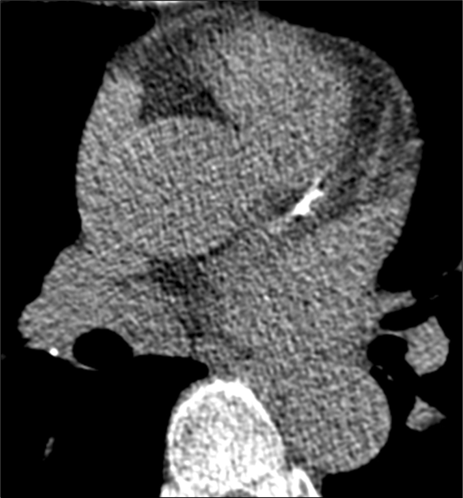 | Fig. 1.Prospective electrocardiography gated computed tomography image of the heart showing calcification in the left coronary artery. |
Table 1.
Demographic, clinical and laboratory characteristics of the 35 patients with systemic sclerosis
| Variable | Total (n=35) | Limited (n=20) | Diffuse (n=15) |
|---|---|---|---|
| Age, years | 52±12 | 52±13 | 50±11 |
| Female (n=33) | 51±12 | ||
| Male (n=2) | 65±4 | ||
| Duration, years | 8±7 | 7±7 | 10±7 |
| BMI, kg/m2 | 22±2 | 21±2 | 22±3 |
| Smoking, pack year | 4±11 | 5±13 | 2±8 |
| Modified Rodnan skin score | 12±7 | 7±4∗ | 19±4∗ |
| Diabetes mellitus, n | 0 | 0 | 0 |
| Hypertension, n | 0 | 0 | 0 |
| Total steroid doses, mg | 1,956±2,838 | 821±1,233† | 3,468±3,634† |
| Six months steroid doses, mg/day | 3±5 | 1±3 | 4±7 |
| Fasting plasma glucose (mg/dl) | 82±14 | 86±11 | 78±17 |
| Total cholesterol, mg/dl | 170±35 | 160±35 | 179±35 |
| HDL, mg/dl | 53±15 | 52±14 | 53±17 |
| LDL, mg/dl | 110±36 | 108±39 | 112±32 |
| Triglyceride, mg/dl | 139±55 | 142±62 | 134±45 |
| CRP, mg/dl | 0.4±0.5 | 0.2±0.3 | 0.5±0.6 |
| ESR, mm/hr | 36±26 | 31±23 | 40±29 |
| ANA, n/total (%) | 34/34 (100) | 20/20 (100) | 14/14 (100) |
| Anti-Scl70 Ab, n/total (%) | 11/34 (32) | 2/20 (10)‡ | 9/14 (64)‡ |
| Anti-centromere Ab, n/total (%) | 4/31 (13) | 3/20 (15) | 1/11 (9) |
| Anti-RNP Ab, n/total (%) | 12/25 (48) | 7/15 (47) | 5/10 (50) |
| Pulmonary involvement, n/total (%) | 28/35 (80) | 16/20 (80) | 12/15 (80) |
| Esophageal involvement, n/total (%) | 30/35 (86) | 18/20 (90) | 12/15 (80) |
Table 2.
Individual characteristics, Framingham risk score and coronary calcification score of the 35 patients with systemic sclerosis
 XML Download