

 PDF
PDF ePub
ePub Citation
 Print
Print


Abstract
Wegener's granulomatosis is a rare disease that pathologically causes necrotizing granulomatous vasculitis in the arterioles and venules and it can invade the whole body. In addition, it is difficult to distinguish between a nontuberculous mycobacteria infection that shows manifestations of granuloma and Wegener's granulomatosis. There has been no reported on a patient who had the 2 abovementioned two diseases at the same time. A 69 year old male patient had Wegener's granulomatosis that had invaded the prostate. He also had a scrotal swelling and back pain. He had manifestations of granulomatous infection on the scrotum and spine biopsies. However, there was no clinical evidence of Wegener's granulomatosis. As a result, we examined him for other diseases that can cause a granuloma. Consequently, he was also diagnosed as suffering with a nontuberculous mycobacteria infection. We report here on this case and we review the relevant medical literature.
References
1. Duna GF, Galperin C, Hoffman GS. Wegener's granulomatosis. Rheum Dis Clin N America. 1995; 21:949–86.

2. Hoffman GS, Specks U. Antineutrophil cytoplasmic antibodies. Arthritis Rheum. 1998; 41:1521–37.
3. Stillwell TJ, DeRemee RA, McDonald TJ, Weiland LH, Engen DE. Prostatic involvement in Wegener's granulomatosis. J Urol. 1987; 138:1251–3.
4. Mundinger A, Pumpe K, Grosser G, Herbst EW, Grotz W, Kropelin T. Primary manifestation of Wegener's granulomatosis of the prostate. Urologe. 1989; 28:234–6.
5. Katalinić-Janković V, Grle SP, Obrovac M, Cvetnić E, Alfirević T. Infections due to nontuberculous mycobacteria. Lijec Vjesn. 2007; 129:146–51.
6. Calabrese LH, Duna G. Vasculitis associated with ant-ineutrophil cytoplasmic antibody. In: Ruddy S, Harris ED, Sledge CB, eds. Kelly's textbook of rheumatology. 2001; 6:1165–84.
7. Cotch MF, Hoffman GS, Yerg DE, Kaufman GI, Tar-gonski P, Kaslow RA. The epidemiology of Wegener's granulomatosis: estimates of the five years period prevalence, annual mortality, and geographic disease distribution from population-based data sources. Arthritis Rheum. 1996; 39:87–92.
8. Leavitt RY, Fauci AS, Bloch DA, Michel BA, Hunder GG, Arend WP, et al. The American college of rheumatology 1990 criteria for the classification of Wegener's granulomatosis. Arthritis Rheum. 1990; 33:1101–7.
9. Morelli S, Gurgo Di Castelmenardo AM, Conti F, Sgreccia A, Alessandri C, Bernardo ML, et al. Cardiac involvement in patients with Wegener's granulomatosis. Rheumatol Int. 2000; 19:209–12.
10. van der Woude FJ, Rasmussen N, Lobatto S, Wiik A, Permin H, van Es LA, et al. Autoantibodies against neutrophils and monocytes: tool for diagnosis and marker of disease activity in Wegener's granulomatosis. Lancet. 1985; 1:425–9.
11. Nolle B, Specks U, Ludemann J, Rohrbach MS, De-Remee RA, Gross WL. Anticytoplasmic autoantibodies: their immunodiagnostic value in Wegener granulomatosis. Ann Intern Med. 1989; 111:28–40.
12. Bray VJ, Hasbargen JA. Prostatic involvement in Wegener's granulomatosis. Am J Kidney Dis. 1991; 17:578–80.
Fig. 1.
(A) The prostate shows a geographically necrotizing granulomatous inflammation. (B) A vasculitis is noted in the small to medium sized vessel. There are many inflammatory cells in the vessel wall and perivascular area (arrow). Fibrinoid necrotic material is noted in the vascular lumen (H&E stain, A ×100, B ×200).


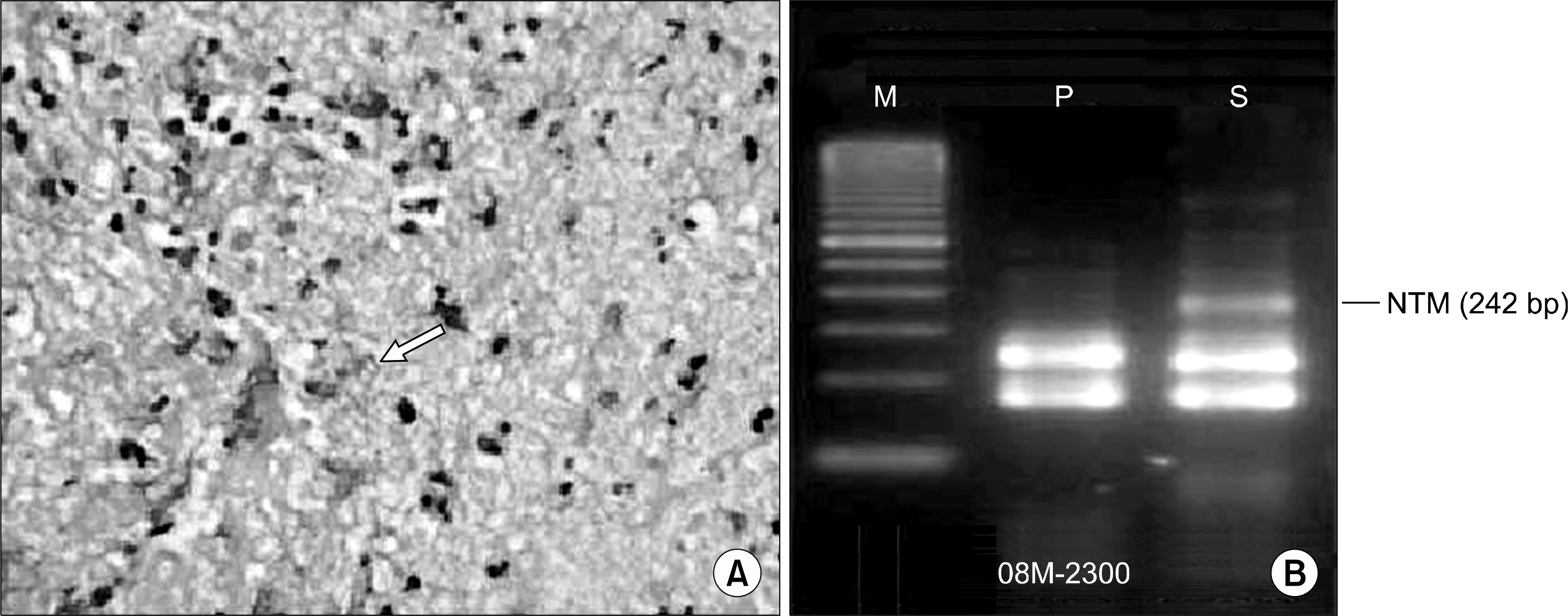
Fig. 3.
(A) An acid fast bacillus (arrow) is noted on the Ziehl-Neelsen stain (A ×1,000), (B) Polymerase chain reaction analysis for NTM. The sample is equal to the result of the positive control (M: mark, P: positive control, S: sample).


 XML Download