

 PDF
PDF ePub
ePub Citation
 Print
Print


Abstract
Wells' syndrome is an inflammatory dermatosis with associated aberrant eosinophil responses caused by unknown factors. Its histology is characterized by erythematous plaques with "flame figures" in the dermis, which is potentially diagnostic but not pathognomic. Cases of Wells' syndrome in patients with Churg-Strauss syndrome (CSS), which is characterized by antineutrophil cytoplasmic antibody-related necrotizing vasculitis, marked peripheral eosinophilia, and eosinophil tissue infiltrates, have rarely been reported, and the pathogenic association between these two diseases remains undetermined. Differences of clinical and histopathologic features of these two diseases suggest that they are distinct disease entities, even though, in part, they share pathogenic mechanisms. Here we present a new case with Wells' syndrome in a patient with CSS, treated with systemic steroid.
References
1. Wells GC. Recurrent granulomatous dermatitis with eosinophilia. Trans St Johns Hosp Dermatol Soc. 1971; 57:46–56.
2. Schuttelaar ML, Jonkman MF. Bullous eosinophilic cellulitis (Wells' syndrome) associated with Churg-Strauss syndrome. J Eur Acad Dermatol Venereol. 2003; 17:91–3.

3. Aberer W, Konrad K, Wolff K. Wells' syndrome is a distinctive disease entity and not a histologic diagnosis. J Am Acad Dermatol. 1988; 18:105–14.
4. Dijkstra JW, Bergfeld WF, Steck WD, Tuthill RJ. Eosinophilic cellulitis associated with urticaria. A report of two cases. J Am Acad Dermatol. 1986; 14:32–8.
5. Schorr WF, Tauscheck AL, Dickson KB, Melski JW. Eosinophilic cellulitis (Wells' syndrome): histologic and clinical features in arthropod bite reactions. J Am Acad Dermatol. 1984; 11:1043–9.
6. Hirsch K, Ludwig RJ, Wolter M, Zollner TM, Hardt K, Kaufmann R, et al. Eosinophilic cellulitis (Wells' syndrome) associated with colon carcinoma. J Dtsch Dermatol Ges. 2005; 3:530–1.
7. 이운하, 최정철, 전덕규. 식도암 환자에서 발생한 Wells 증후군 1예. 대한피부과학회지. 2000; 43:1130–2.
8. Lee SC, Shin SS, Lee JB, Won YH. Wells syndrome associated with Churg-Strauss syndrome. J Am Acad Dermatol. 2000; 43:556–7.
9. Govoni M, Colina M, Cavazzini L, Trotta F. Churg-Strauss syndrome and Wells syndrome: coincidence or pathogenetic association? A new case report. Clin Exp Rheumatol. 2007; 25(Supple 44):41.
10. Masi AT, Hunder GG, Lie JT, Michel BA, Bloch DA, Arend WP, et al. The American College of Rheumatology 1990 criteria for the classification of Churg-Strauss syndrome. Arthritis Rheum. 1990; 33:1094–100.
11. Pagnoux C, Guilpain P, Guillevin L. Churg-Strauss syndrome. Curr Opin Rheumatol. 2007; 19:25–32.
12. Strauss L, Churg J, Zak FG. Cutaneous lesions of allergic granulomatosis; a histopathologic study. J Invest Dermatol. 1951; 17:349–59.
13. Herr H, Koh JK. Eosinophilic cellulitis (Wells' syndrome) successfully treated with low-dose cyclosporine. J Korean Med Sci. 2001; 16:664–8.
14. 이정훈, 전희대, 이성렬, 박영립, 이종석, 황규왕. 전신 PUVA로 치료한 Wells 증후군 1예. 대한피부과학회 지. 2000; 38:949–54.
15. 김은경, 박찬금, 이중달. 호산구성 봉와직염의 임상 및 병리 조직학적 연구. 대한병리학회지. 1995; 29:334–42.
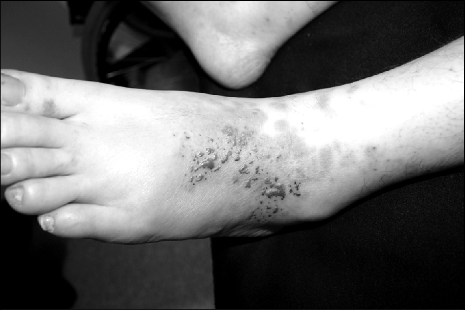
Fig. 2.
There is a heavy dermal infiltration of inflammatory cells (A: H&E, ×40), composed of lymphocytes and histiocytes with conspicuous eosinophils (B: H&E, ×200).

Fig. 3.
Among dermal collagen bundles, there are collections of eosinophils with eosinophilic granules, so called "flame figures" (H&E, ×400).
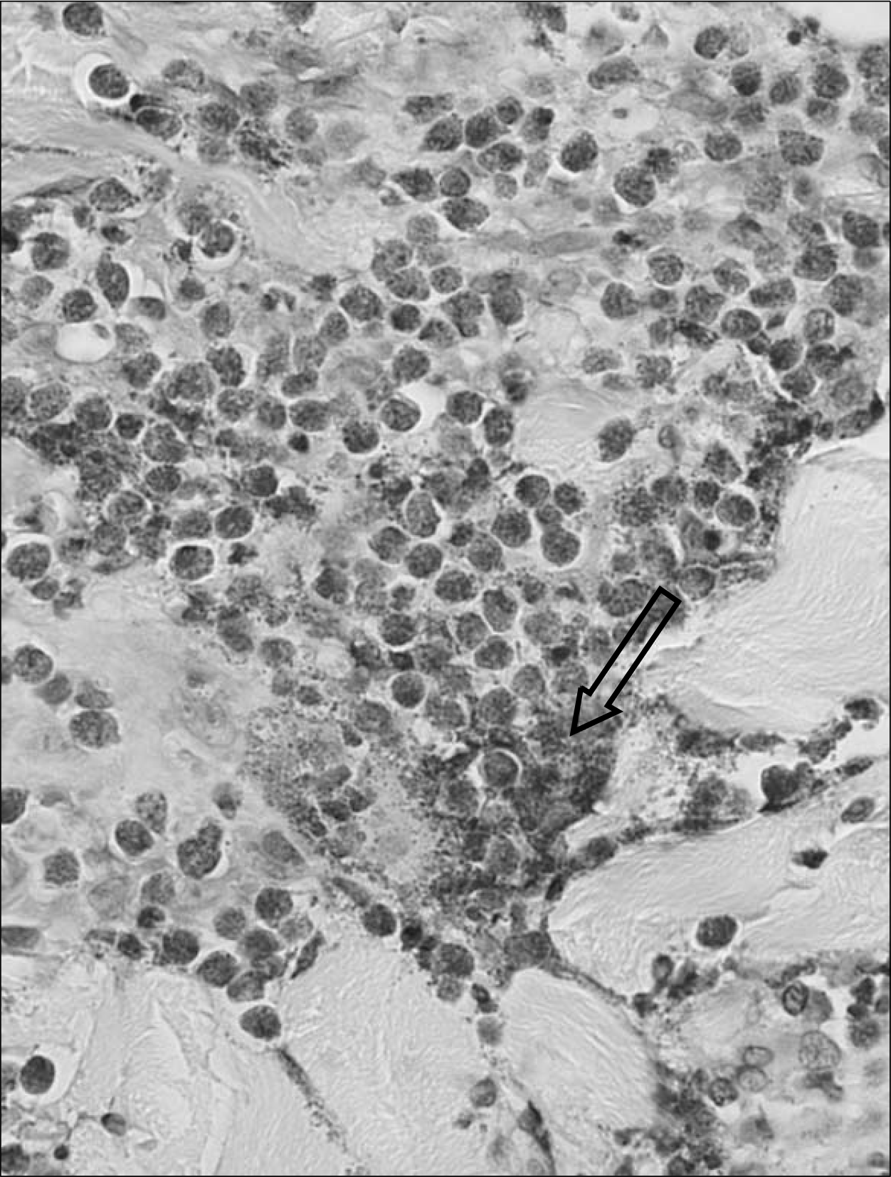
Table 1.
Characteristics in Wells' syndrome with Churg-Strauss syndrome
 XML Download