Abstract
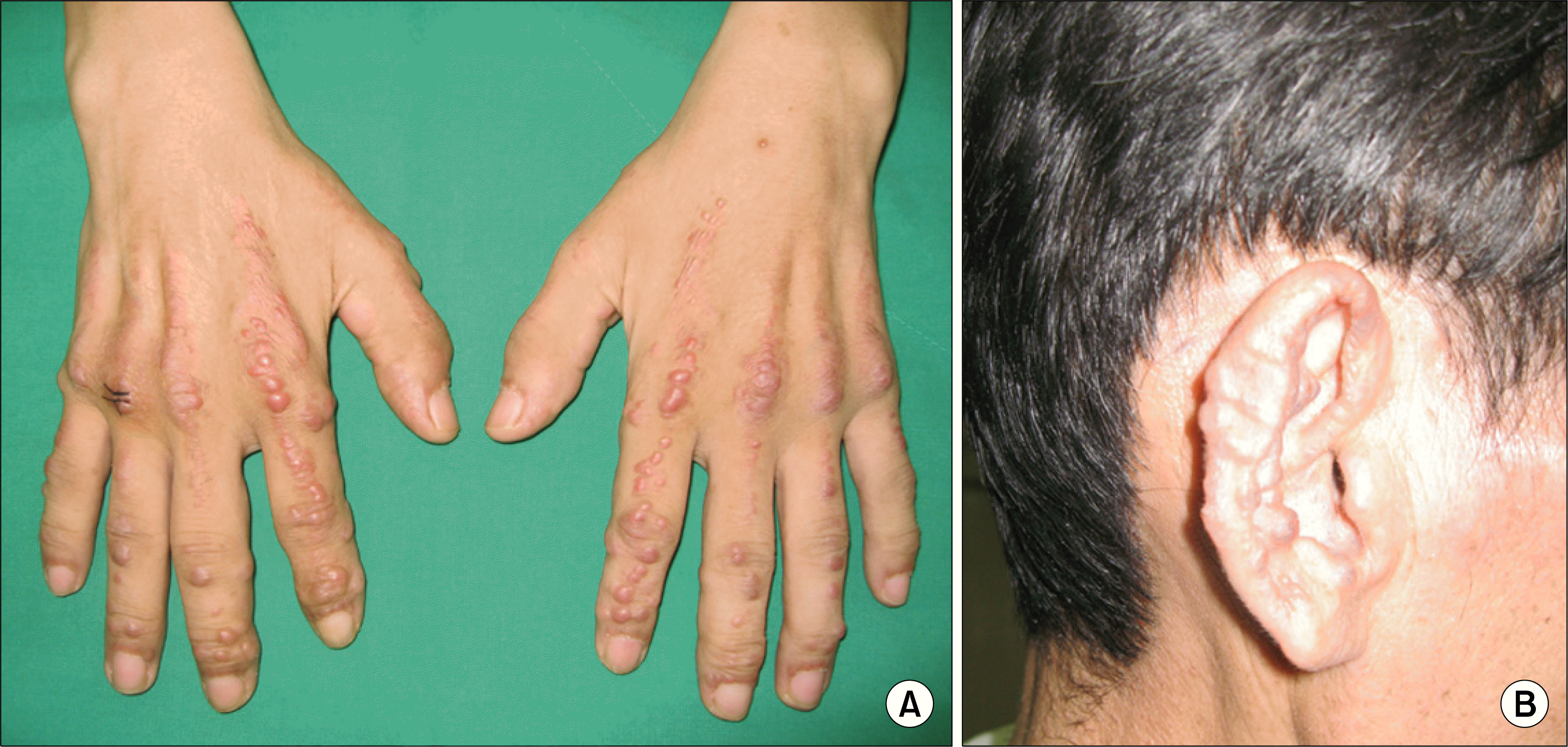
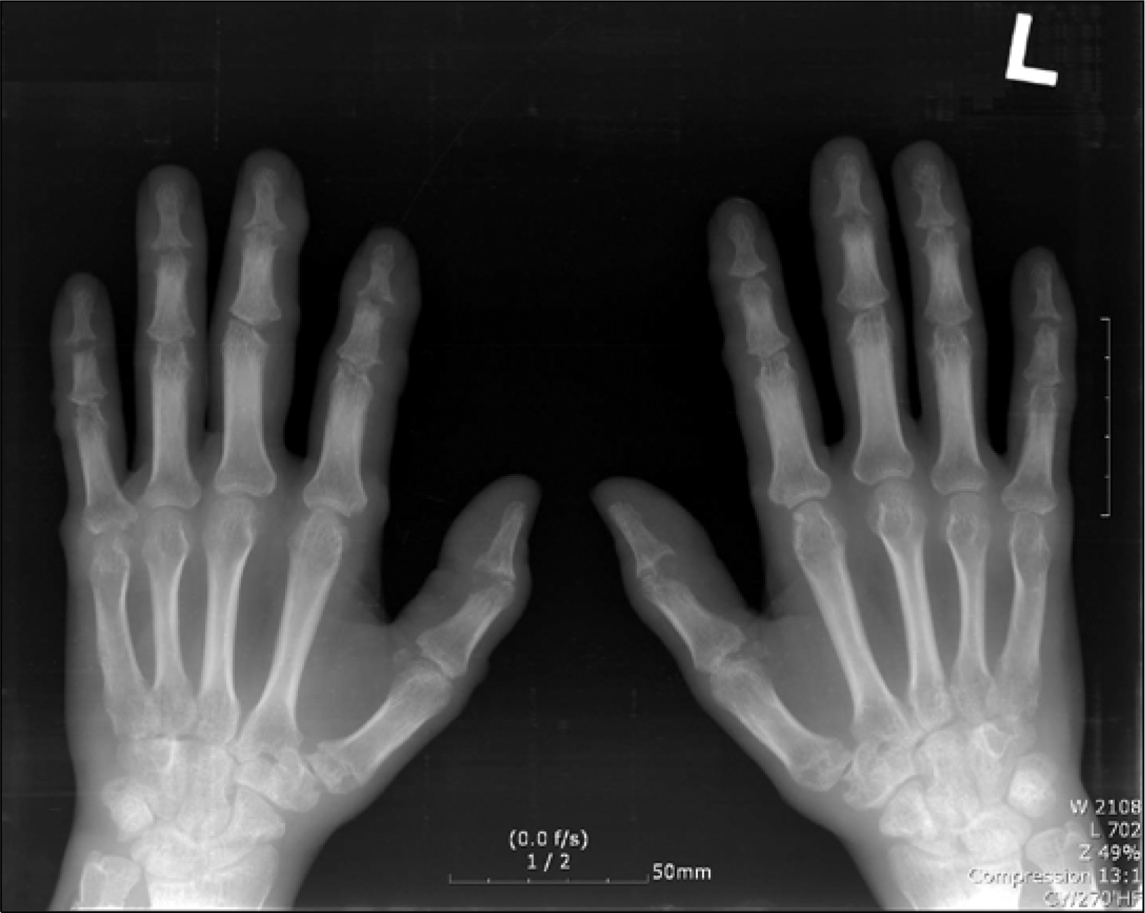
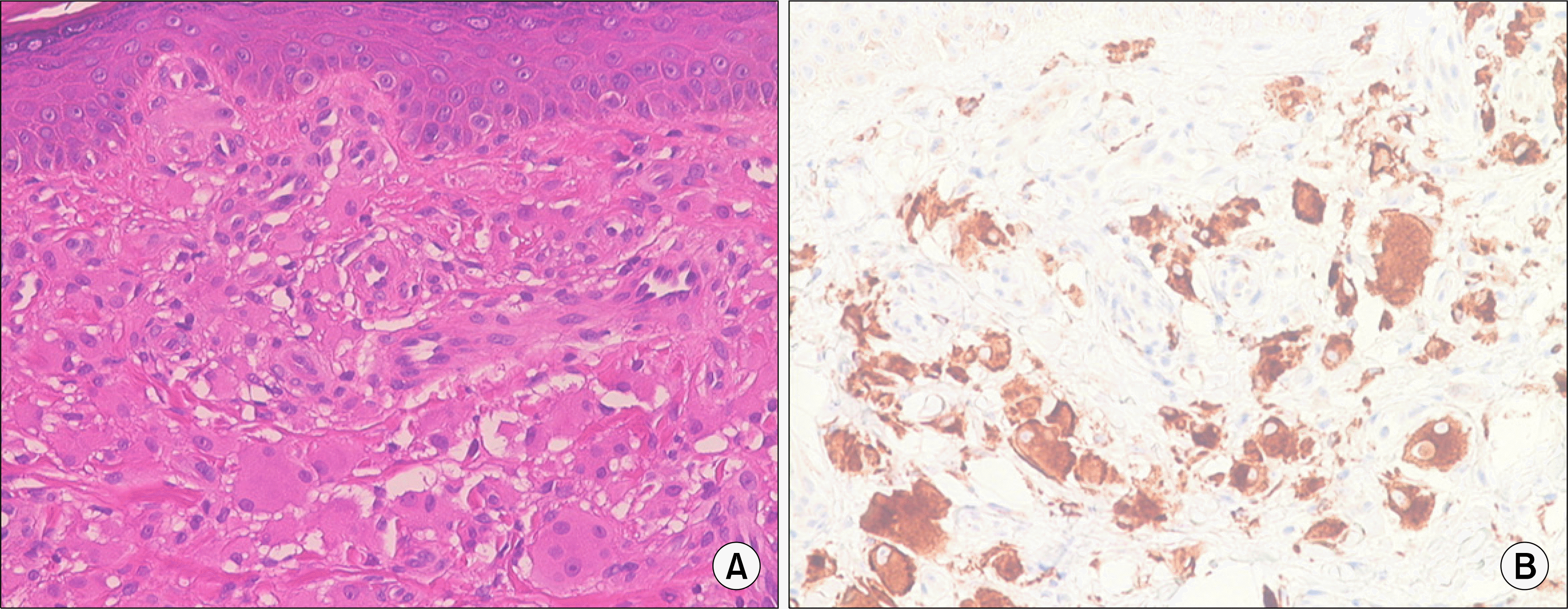
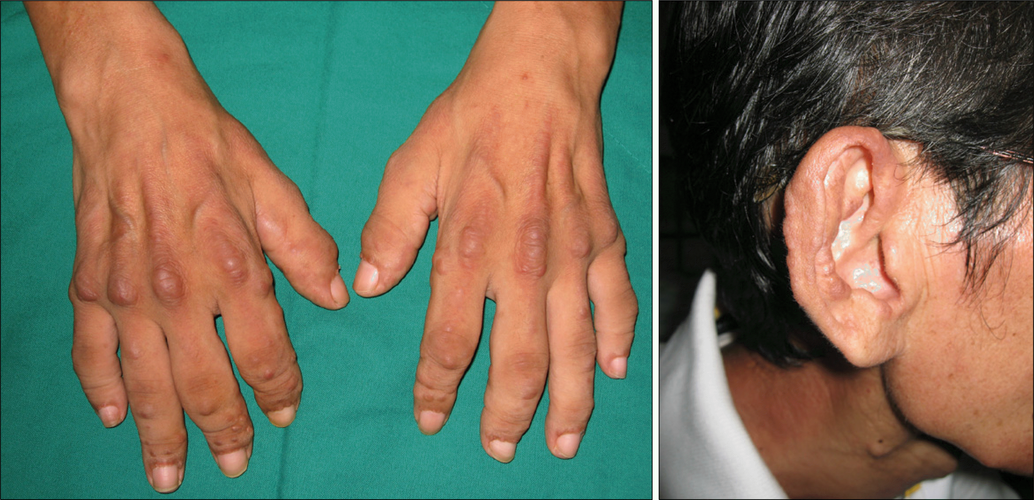
Multicentric reticulohistiocytosis (MRH) is a rare disease affecting joints, skin and internal organs. Yet the cause is still unknown. In most cases, it can be misdiagnosed as rheumatoid arthritis or psoriatic arthritis before typical change in the skin emerges, and precise diagnosis is essential because it may become severe enough to transform and destroy the joints. Therefore, skin or synovial biopsy can confirm the existence of this disease. This particular patient is a 39-year-old male who had been treated for rheumatoid arthritis on a wrist and hand and transferred to this hospital when arthritic pain continued with erythematous papules and nodules on the hands, the outer rims of the ears, hands and elbows. The X-ray examination of both hands revealed multiple marginal erosion of proximal and distal interphalangeal joints and destruction of subchondral bones. periarticular osteoporosis and joint space enlargement is combined, but no new bones were seen to be formed. Biopsy of erythematous nodules on the dorsum of hands showed that an infiltrate histiocytes and multi-nuclear giant cells were aligned irregularly, and immunological chemical staining showed potential for being positive to PAS and CD68. To control pain and regulate activity of the disease, tracing observation and treatment were started from the outside with NSAID, hydroxycholoquine, MTX and prednisolone. A month of treatment did not improve arthritis and skin problems, and increased dose of MTX, prednisolone did improve arthritis a little but not skin problems. Treatment with infliximab (3
mg/kg), a anti-tumor necrosis factor, is in progress, showing improvement in both conditions.
References
1. Katerine AC, Tomas MR, Jag B, Alpdogan K, Eugene YK. Multicentric Reticulohistiocytosis: a systemic osteoclastic Disease? Arthritis Rheum. 2008; 59:444–8.

2. Mohammad BO, Golbarg M, Hossein S. Multicentric reticulohistiocytosis. APLAR J Rheumatol. 2007; 10:330–2.
3. Jennifer DG, Carol DH, Ralph S, John HK, John CD. Multicentric reticulohistiocytosis. Arthritis Rheum. 2000; 43:930–8.
4. Santilli D, Monaco AL, Cacazzini PL, Trotta F. Multicentric reticulohistiocytosis: a rare cause of erosive arthropathy of the distal interphalangeal finger joints. Ann Rheum Dis. 2002; 61:485–7.

5. Tajirian AL, Mohsin KM, Leslie RB, Edward VL. Multicentric reticulohistiocytosis. Clin Dermatol. 2006; 24:486–92.

6. Kang JH, Lee CW, Sung YK, Bae SC. A case of multicentric reticulohistiocytosis. Korean J Dermatol. 2005; 43:672–4.
7. Shahram B, Farrokh K, Mohsen DZ, Abdol AM. Multicentric reticulohistiocytosis presenting with papulonodular skin eruption and polyarthritis. Eur J Dermatol. 2005; 15:196–200.
8. Kim KH, Choi SW, Lee SH, Lee SW, Chung WT, Kim DC, et al. A case of multicentric reticulohistiocytosis. Korean J Med 67;. 850–6.
9. Bradley TK, Kenneth TC, John SW, William WG. Treatment of multicentric reticulohistiocytosis with etanercept. Arch Dermatol. 2004; 140:919–21.

10. Lambert CM, Nuki G. Multicentric reticulohistiocytosis with arthritis and cardiac infiltration: regression following treatment for underlying malignanacy. Ann Rheum dis. 1992; 51:815–7.
11. Sokka T, Pincus T. Quantitative joint assessment in rheumatoid arthritis. Clin Exp Rheumatol. 2005; 23(Suppl 39):58–62S.
Fig. 1.
Erythematous papulonodular skin lesion on the dorsum of the hands (A) and around the auricle (B).

Fig. 2.
Both hand AP show the presence of multiple marginal erosions and subchondral bone destruction of the all DIP and PIP joints and first MP joint. Multiple soft tissur nodules are also detected on these joints. Joints spaces are enlarged and the new bone formation is not seen. Periarticular osteoporosis is combined.


 PDF
PDF ePub
ePub Citation
 Print
Print


 XML Download