

 PDF
PDF ePub
ePub Citation
 Print
Print


Abstract
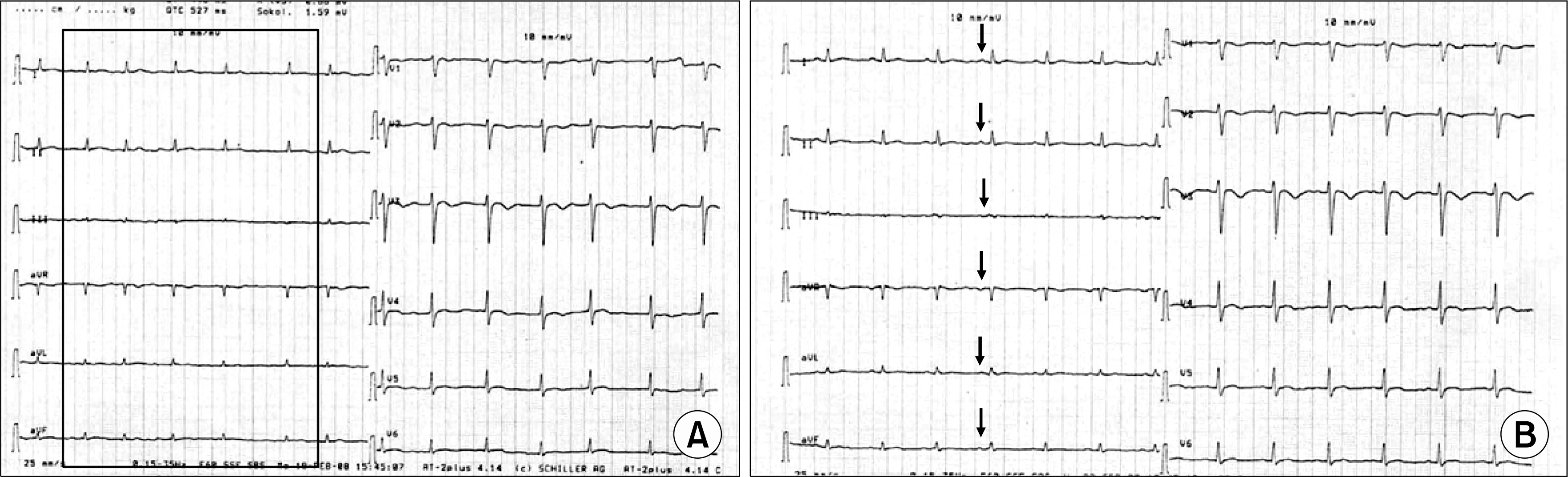
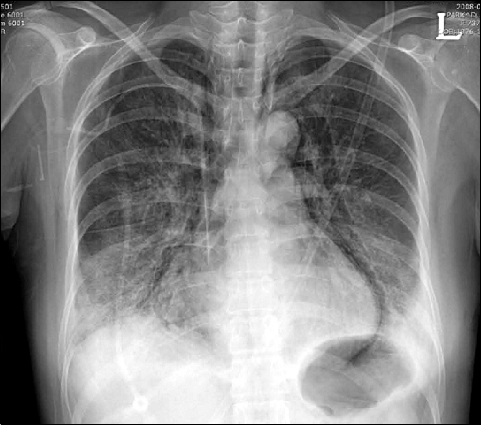
Systemic lupus erythematous (SLE) is systemic autoimmune disease of unknown etiology. SLE involve all part of heart but clinically important myocarditis is an unusual feature and is potentially fatal complication of SLE. We describe the woman who had diagnosed as SLE, 5 years ago and in that time, she had sinus tachycardia, mild dyspnea, chest discomfort, and depressed left ventricular function. She was diagnosed to myocarditis clinically and was treated by high-dose corticosteroids of intravenous pulse methylprednisolone. After treatment, she improved and showed improvement of left ventricular function. However, a sudden pneumomediastinum was occurred without trauma, and she died. The cause of pneumomediastinum was unexplained. We report a sudden pneumomediastinum in the course of lupus myocarditis presenting as sinus tachycardia with review of literature.
References
1. Seferovic PM, Ristic AD, Maksimovic DS, Sime-unovic G, Ristic G, Radovanovic D, et al. Cardiac arrhythmias and conduction disturbances in autoimmune rheumatic diseases. Rheumatology (oxford). 2006; 45(Suppl):39–42S.
2. D'Cruz D, Khamashta M, Huges GRV. Cardiovascular manifestation of systemic lupus erythematousus. Dubois' lupus erythematous. 6th ed.p. 645, Philadelphia, Lippincott Williams and Wilkims;2002.
3. Wijetunga M, Rockson S. Myocarditis in systemic lupus erythematosus. Am J Med. 2002; 113:419–42.

4. Moder KG, Miller TD, Tazelaar HD. Cardiac involvement in systemic lupus erythematosus. Mayo Clin Proc. 1999; 74:275–84.
5. Guzman J, Cardiel MH, Arce-Salinas A, Alarcon-Segovia D. The contribution of resting heart rate and routine blood tests to the clinical assessment of disease activity in systemic lupus erythematosus. J Rheumatol. 1994; 21:1845–8.
6. Cheug SM, Chang DM, Lee WH, Ding Ya. Acute myocarditis as an initial manifestation of systemic lupus erythromatosus: a case report. Chung Hua I Hsueh Tsa Chih (Taipei). 1996; 58:205–8.
7. Frustaci A, Gentiloni N, Caldarulo M. Acute myocarditis and left ventricular aneurysm as presentations of systemic lupus erythematosus. Chest. 1996; 109:282–4.
8. Logar D, Kveder T, Rozman B, Dovovisek J. Possible association between anti-Ro antibodies and myocarditis or cardiac conduction defects in adults with systemic lupus erythematosus. Ann Rheum Dis. 1990; 49:627–9.
9. Comin-Colet J, Sachez-Corral MA, Alegre-Sancho JJ, Valverde J, Lopez-Gomez D, Sabate X, et al. Complete heart block in an adult with systemic lupus erythematosus and recent onset of hydroxychloroquine therapy. Lupus. 2001; 10:59–62.
10. Law WG, Thong BY, Lian TY, Kong KO, Chng HH. Acute lupus myocarditis: clinical features and outcome of an oriental series. Lupus. 2005; 14:827–31.
11. Feldman AM, McNamara D. Myocarditis. N Engl J Med. 2000; 343:1388–98.
12. Doherty NE III, Feldman G, Maurer G, Siegel RJ. Echocardiographic findings in systemic lupus erythematosus. Am J Cardiol. 1988; 61:1144.
13. Chan YK, Li EK, Tam LS, Chow LT, Ng HK. Intravenous cyclophosphamide improves cardiac dysfunction in lupus myocarditis. Scand J Rheumatol. 2003; 32:306–8.
14. Doria A, Iaccarino L, Sarzi-Puttini P, Atzeni F, Turriel M, Petri M. Cardiac involvement in systemic lupus erythematosus. Lupus. 2005; 14:683–6.
15. Paira SO, Roverano S. Bilateral pneumothorax and mediastinal emphysema in systemic lupus erythematosus. Clin Rheumatol. 1992; 11:571–3.
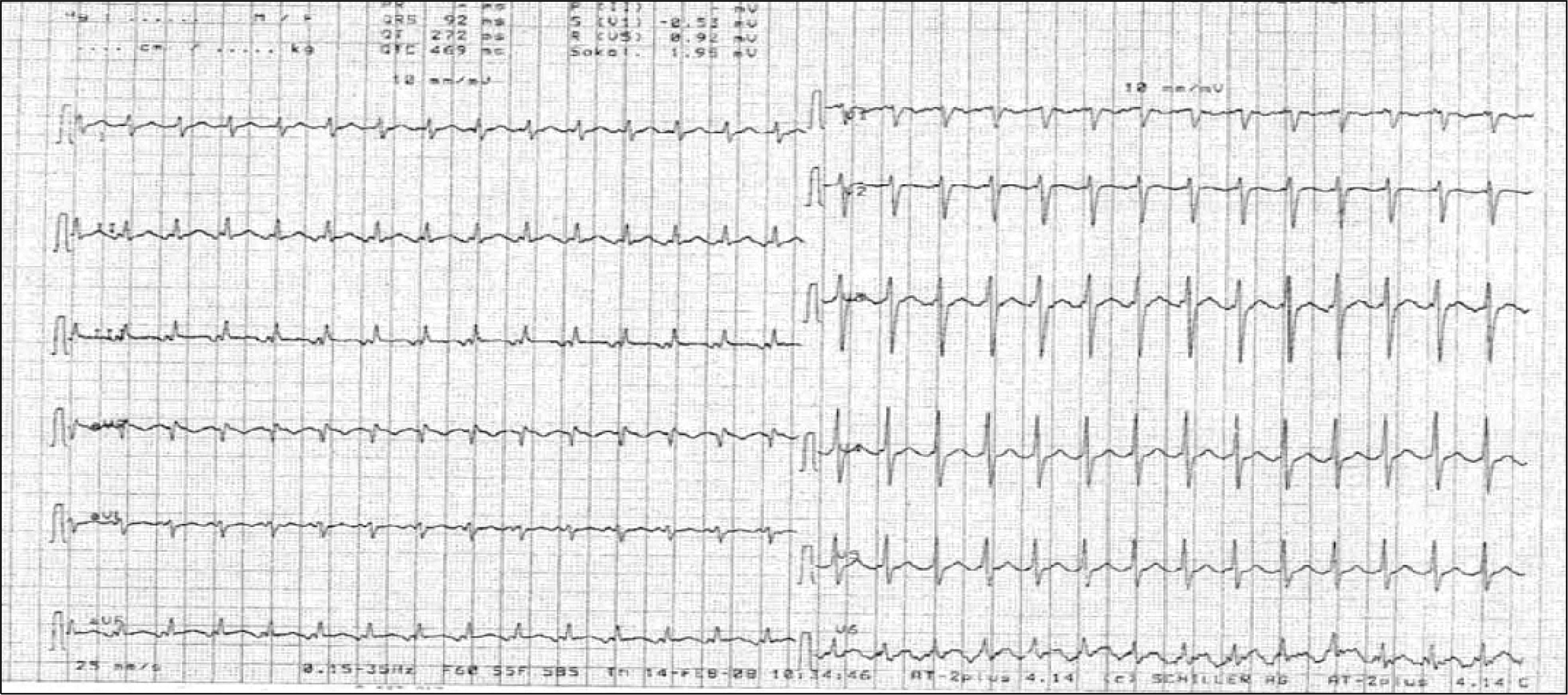
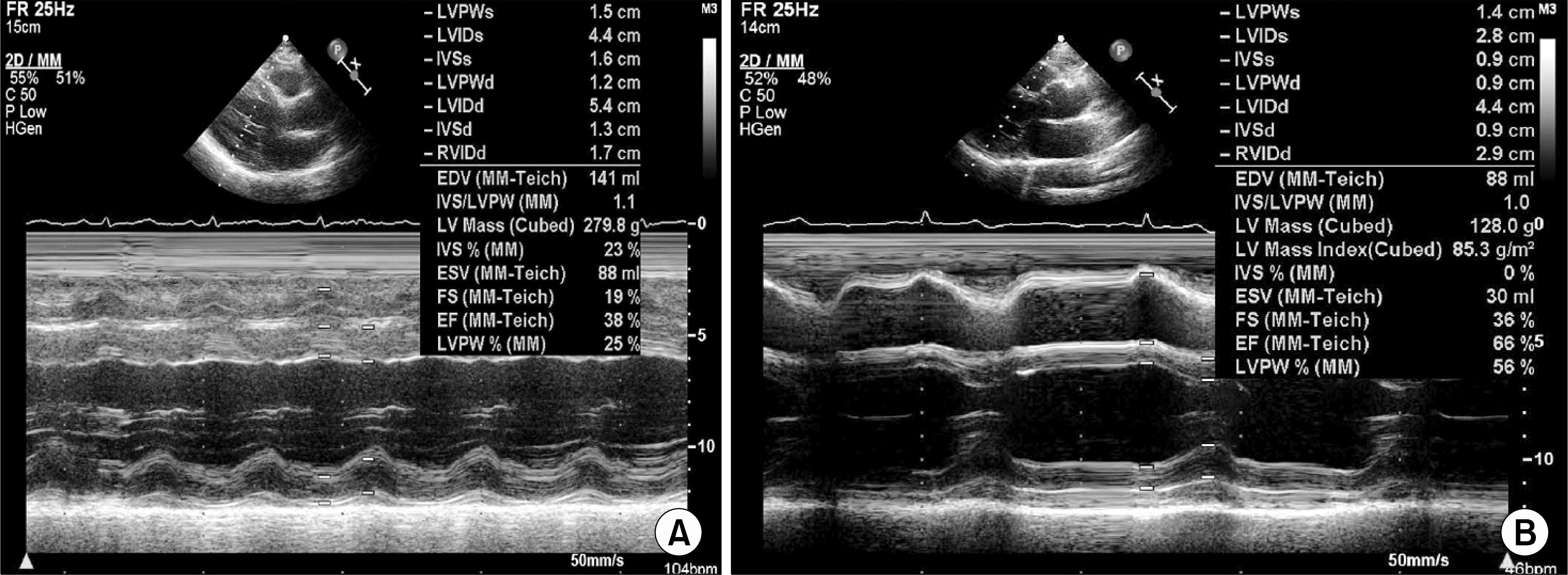
Fig. 2.
Transthoracic echocardiography (M-mode echocadiography) shows decreased ejection fraction (38%) (A). Follow-up transthoracic echocardiography shows improved ejection fraction (66%) than before (38%) (B).
 XML Download