Abstract
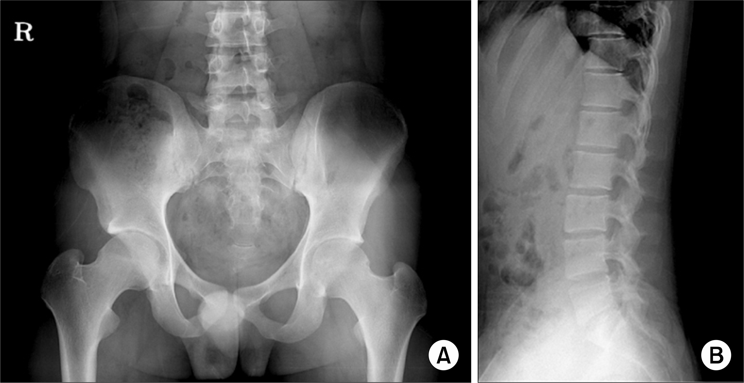
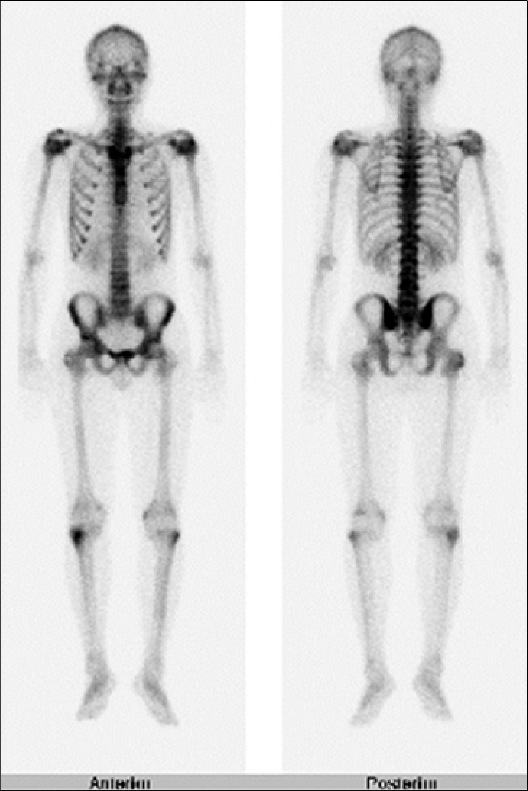
Ankylosing spondylitis (AS) is a common inflammatory arthritis that affects the axial skeleton, causing characteristic inflammatory back pain, which can lead to structural and functional impairments and a decrease in quality of life. The disease can be accompanied by extraskeletal manifestations, such as acute anterior uveitis, inflammation in the colon or ileum, aortic insufficiency, cardiac conduction defects, fibrosis of the upper lobes of the lungs, neurologic involvement, or renal (secondary) amyloidosis. We report the case of a 19 year-old man who developed Henoch-Schönlein purpura (HSP) and subsequently AS. It has been recognized that AS may be associated with cutaneous vasculitis and IgA nephropathy, but the association of HSP with AS has not been reported. This association of IgA nephropathy or HSP with AS raises the possibility of a common or related pathogenesis.
References
2. Kasper DL, Braunwald E, Fauci AS, Hauser SL, Longo DL, Jameson JL, et al. Harrison's principles of internal medicine. 16th ed. p.1993–4. New York, McGraw-Hill,. 2005.
3. Jennette JC, Ferguson AL, Moore MA, Freeman DG. IgA nephropathy associated with seronegative spondylarthropathies. Arthritis Rheum. 1982; 25:144–9.
4. Peeters AJ, van den Wall Bake AW, Daha MR, Breedveld FC. Inflammatory bowel disease and ankylosing spondylitis associated with cutaneous vasculitis, glomerulonephritis, and circulating IgA immune complexes. Ann Rheum Dis. 1990; 49:638–40.

5. Beauvais C, Kaplan G, Mougenot B, Michel C, Marinho E. Cutaneous vasculitis and IgA glomerulonephritis in ankylosing spondylitis. Ann Rheum Dis. 1993; 52:61–2.

6. Hsu CM, Kuo SY, Chu SJ, Shih TY, Chen A, Huang GS, Chang DM, et al. Coexisting IgA nephropathy and leukocytoclastic cutaneous vasculitis associated with ankylosing spondylitis: a case report. Zhonghua Yi Xue Za Zhi. 1995; 55:83–8.
8. Cowling P, Ebringer R, Ebringer A. Association of inflammation with raised serum IgA in ankylosing spondylitis. Ann Rheum Dis. 1980; 39:545–9.

9. Deicher H, Ebringer A, Hildebrand S, Kemper A, Zeidler H. Circulating immune complexes in ankylosing spondylitis. Br J Rheumatol. 1983; 22:122–7.

10. Reynolds TL, Khan MA, van der Linden S, Cleveland RP. Differences in HLA-B27 positive and negative patients with ankylosing spondylitis: study of clinical disease activity and concentrations of serum IgA, C reactive protein, and haptoglobin. Ann Rheum Dis. 1991; 50:154–7.

11. Collado A, Sanmarti R, Bielsa I, Castel T, Kanter-ewicz E, Canete JD, et al. Immunoglobulin A in the skin of patients with ankylosing spondylitis. Ann Rheum Dis. 1988; 47:1004–7.

12. van de Laar MA, Moens HJ, van der Korst JK. Absence of an association between ankylosing spondylitis and IgA nephropathy. Ann Rheum Dis. 1989; 48:262–4.

13. Rao JK, Allen NB, Pincus T. Limitation of the 1990 American College of Rheumatology classification criteria in the diagnosis of vasculitis. Ann Intern Med. 1998; 129:345–52.
14. Davin JC, Weeing JJ. Diagnosis of Henoch-Schönlein purpura: renal or skin biopsy? Pediatr Nephrol. 2003; 18:1201–3.

15. Shrestha S, Sumingan N, Tan J, Althous H, McWill-iam L, Ballardie F, et al. Henoch Schönlein purpura with nephritis in adults: adverse prognostic indicators in a UK population. Q J Med. 2006; 99:253–65.
Fig. 1.
(A) Glomeruli show mild mesangiopathy with thin capillary walls. Tubules contain RBC casts (Jones' Silver stain, ×200). (B) There is mild mesangial proliferation with fuchsinophilic deposits (Masson's trichrome stain, ×400). (C) Immunofluorescence staining reveals diffuse mesangial deposits for IgA (×200).


 PDF
PDF ePub
ePub Citation
 Print
Print


 XML Download