

 PDF
PDF ePub
ePub Citation
 Print
Print


References
1. Constantinou M, Vicenzino B. Differential diagnosis of a soft tissue mass in the calf. J Orthop Sports Phys Ther. 2005; 35:88–94.

2. Kane D, Balint PV, Gibney R, Bresnihan B, Sturrock RD. Differential diagnosis of calf pain with musculoskeletal ultrasound imaging. Ann Rheum Dis. 2004; 63:11–4.
3. Jamadar DA, Jacobson JA, Theisen SE, Theisen SE, Ebrahim F, Kalume-Brigido M. Sonography of the painful calf: differential considerations. Am J Roentgenol. 2002; 179:709–16.
4. Sato O, Kondoh K, Iyori K, Kimura H. Midcalf ultrasonography for the diagnosis of ruptured Baker's cysts. Surg Today. 2001; 31:410–3.
5. Khan SY, Hilton-Jones D, Rigby SP. A swollen calf. Lancet. 2005; 365:1662.
6. Cho KH, Lee DY, Kim CW. Erythema induratum of Bazin. Int J Dermatol. 1996; 35:802–8.
7. Khellaf M, Hamidou M, Pagnoux C, Michel M, Brisseau JM, Chevallier X, et al. Vasculitis restricted to the lower limbs: a clinical and histopathological study. Ann Rheum Dis. 2006; 66:554–6.
8. Christopoulos C, Savva S, Pylarinou S, Diakakis A, Papavassiliou E, Economopoulos P. Localised gastrocnemius myositis in Crohn's disease. Clin Rheumatol. 2003; 22:143–5.
9. Gaitini D. Current approaches and controversial issues in the diagnosis of deep vein thrombosis via duplex Doppler ultrasound. J Clin Ultrasound. 2006; 34:289–7.
10. Wigley RD. Popliteal cysts: variations on a theme of Baker. Semin Arthritis Rheum. 1982; 12:1–10.
11. Handy JR. Popliteal cysts in adults: a review. Semin Arthritis Rheum. 2001; 31:108–8.
12. Scott WN, Jacobs B, Lockshin MD. Posterior compartment syndrome resulting from a dissecting popliteal cyst. Clin Orthop. 1977; 122:189–92.
13. Nakano KK. Entrapment neuropathy from Baker's cyst. JAMA. 1978; 239:135.
14. Tashjian RZ, Nickisch F, Dennison D. Ruptured septic popliteal cyst associated with psoriatic arthritis. Orthopedics. 2004; 27:231–3.
15. Fang CS, McCarthy CL, McNally EG. Intramuscular dissection of Baker's cysts: report on three cases. Skeletal Radiol. 2004; 33:367–71.
16. Volteas SK, Labropoulos N, Leon M, Kalodiki E, Nicolaides AN. Incidence of ruptured Baker's cyst among patients with symptoms of deep vein thrombosis. Br J Surg. 1997; 84:342.
17. Langsfeld M, Matteson B, Johnson W, Wascher D, Goodnough J, Weinstein E. Baker's cysts mimicking the symptoms of deep vein thrombosis: diagnosis with venous duplex scanning. J Vasc Surg. 1997; 25:658–62.
18. Fernando RA, Somers S, Edmonson RD, Sidhu PS. Subcutaneous fat necrosis: hypoechoic appearance on sonography. J Ultrasound Med. 2003; 22:1387–90.
19. Hench PK, Reid RT, Reames PM. Dissecting popliteal cyst simulating thrombophlebitis. Ann Int Med. 1966; 64:1259–64.
20. Kilcoyne RF, Imray TJ, Stewart ET. Ruptured Baker's cyst simulating acute thrombophlebitis. JAMA. 1978; 240:1517–8.
21. Katz RS, Zizic TM, Arnold WP, Stevens MB. The pseudothrombophlebitis syndrome. Medicine. 1977; 56:151–64.
22. Liebling MR. Editorial response: Why a duck?–Or for that matter, why a cyst? Clin Infect Dis. 1999; 29:279–80.
23. Tashjian RZ, Nickisch F, Dennison D. Ruptured septic popliteal cyst associated with psoriatic arthritis. Orthopedics. 2004; 27:231–3.
24. Ozgocmen S, Kaya A, Kocakoc E, Kamanli A, Ardicoglu O, Ozkurt-Zengin F. Rupture of Baker's cyst producing pseudothrombophlebitis in a patient with Reiter's syndrome. Kaohsiung J Med Sci. 2004; 27:231–3.
25. Reilly PA, Maddison PJ. Painful, swollen calf in a patient with SLE. Br J Rheumatol. 1987; 26:319–20.
26. Miller TT, Staron RB, Koenigsberg T, Levin TL, Feldman F. MR imaging of Baker cysts: association with internal derangement, effusion, and degenerative arthropathy. Radiology. 1996; 201:247–50.
27. Marti-Bonmati L, Molla E, Dosda R, Casillas C, Ferrer P. MR imaging of Baker cysts −prevalence and relation to internal derangements of the knee. MAGMA. 2000; 10:205–10.
28. Rupp S, Seil R, Jochum P, Kohn D. Popliteal cysts in adults. Prevalence, associated intraarticular lesions, and results after arthroscopic treatment. Am J Sports Med. 2000; 30:112–5.
29. Dirschl DR, Lachiewicz PF. Dissecting popliteal cyst as the presenting symptom of a malfunctioning total knee arthroplasty. Report of four cases. J Arthroplasty. 1992; 7:37–41.
30. von Schroeder HP, Ameli FM, Piazza D, Lossing AG. Ruptured Baker's cyst causes ecchymosis of the foot. A differential clinical sign. J Bone Joint Surg Br. 1993; 75:316–7.
31. Simon HE, Sacchet HA. Muscle hernias of the leg. Review of literature and report of twelve cases. Am J Surg. 1945; 67:87–9.
32. Braunstein JT, Crues JV 3rd. Magnetic resonance imaging of hereditary hernias of the peroneus longus muscle. Skeletal Radiol. 1995; 24:601–4.
33. Sherry RH. Herniation of peroneus brevis muscle: report of a case. Bull Hosp Dis. 1942; 3:69–72.
34. Alhadeff J, Lee CK. Gastrocnemius muscle herniation at the knee causing peroneal nerve compression resembling sciatica. Spine. 1995; 20:612–4.
35. Beggs I. Sonography of muscle hernias. Am J Roentgenol. 2003; 180:395–9.
36. Morris SJ, Adams H. Case report: paediatric intramuscular haemangiomata-don't overlook the phlebolith! Br J Radiol. 1995; 68:208–11.
37. Bloem JL. Imaging of soft tissue tumors. J Belge Radiol. 1992; 75:265–73.
38. Sidhu PS, Rich PM. Sonographic detection and characterization of musculoskeletal and subcutaneous tissue abnormalities in sickle cell disease. Br J Radiol. 1999; 72:9–17.
39. Hung GD, Chen YH, Chen DY, Lan JL. Subcutaneous panniculitis-like T-cell lymphoma presenting with hemophagocytic lymphohistiocytosis and skin lesions with characteristic high-resolution ultrasonographic findings. Clin Rheumatol. 2007; 26:775–8.
40. Tillman C, Holst R, Svedman C. Traumatic fat necrosis: a case report. Acta Derm Venereol. 2003; 83:227–8.
41. Guo M, Lemos L, Baliga M. Nodular sarcoid myositis of skeletal muscle diagnosed by fine needle aspiration biopsy. A case report. Acta Cytol. 1999; 43:1171–6.
Fig. 1.
A 46-year-old woman with rheumatoid arthritis and ruptured and dissected baker cyst. Ultrasonogram shows typical location of the baker cyst in the space between the medial head of the gastrocnemius and semembranosus (A). Anechoic dissected baker cyst with internal synovial proliferation demonstrates in the space between the medial head gastrocneiums and superficial fascia (B). Coronal fat suppressed T2-weighted MR image show high signal intensity of baker cyst with rupture and dissection into the space between the medial head gastrocnemius and superficial fascia. There is an extension into the subcutaneous tissue layer (C, arrow).
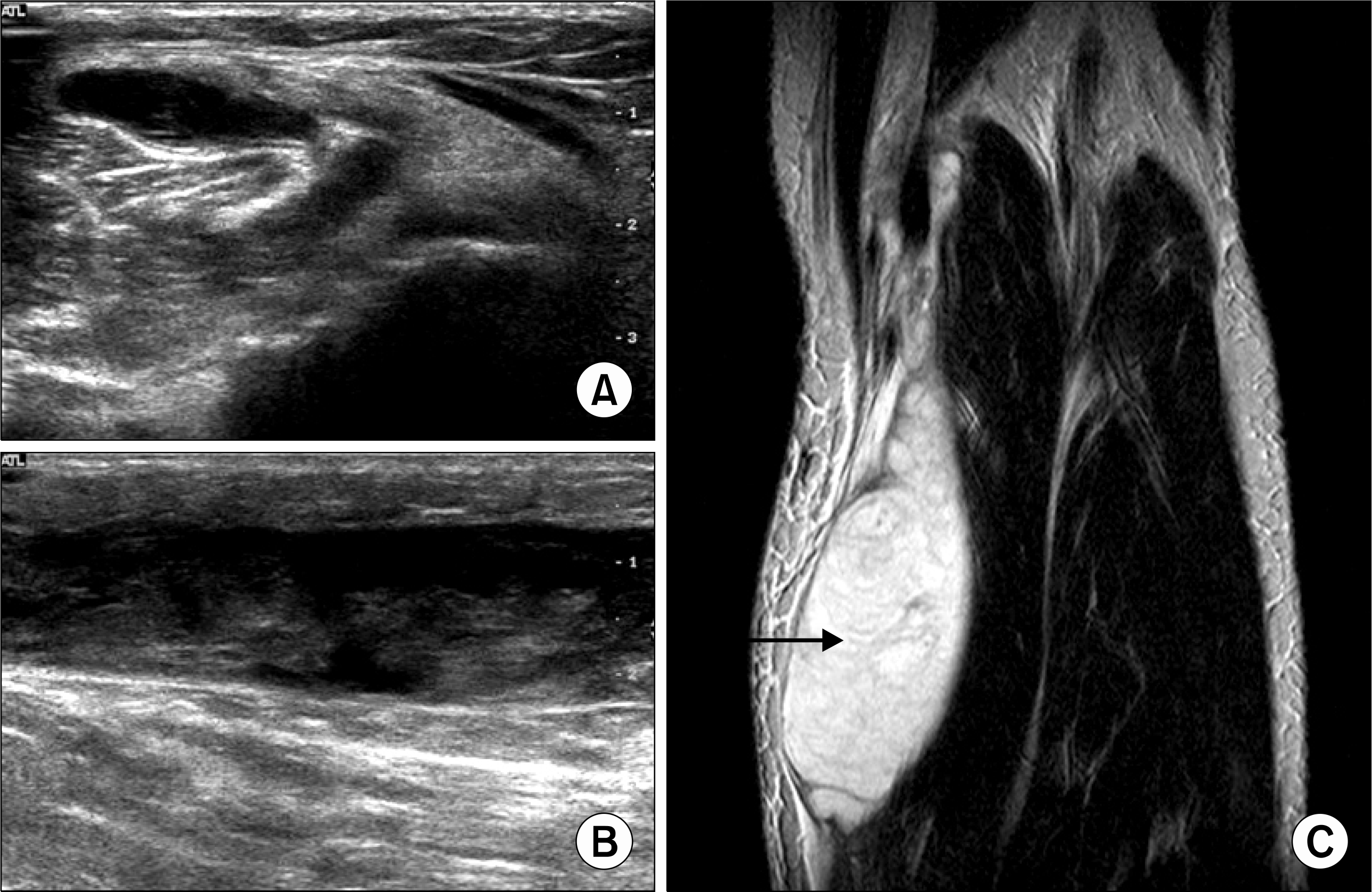
Fig. 2.
A-72-year old woman with rheumatoid arthritis and ruptured and dissected baker cyst. Axial T1-weighted MR image (A), T2-weighted image (B), and coronal sagittal fat-suppressed image (C) show ruptured and dissected baker cyst, appearing with heterogeneous high signal intensity due to internal synovial proliferation and hemorrhage within the gastrocnemius muscle and space between the superficial fascia and the both head of the gastrocnemius muscle (arrow).
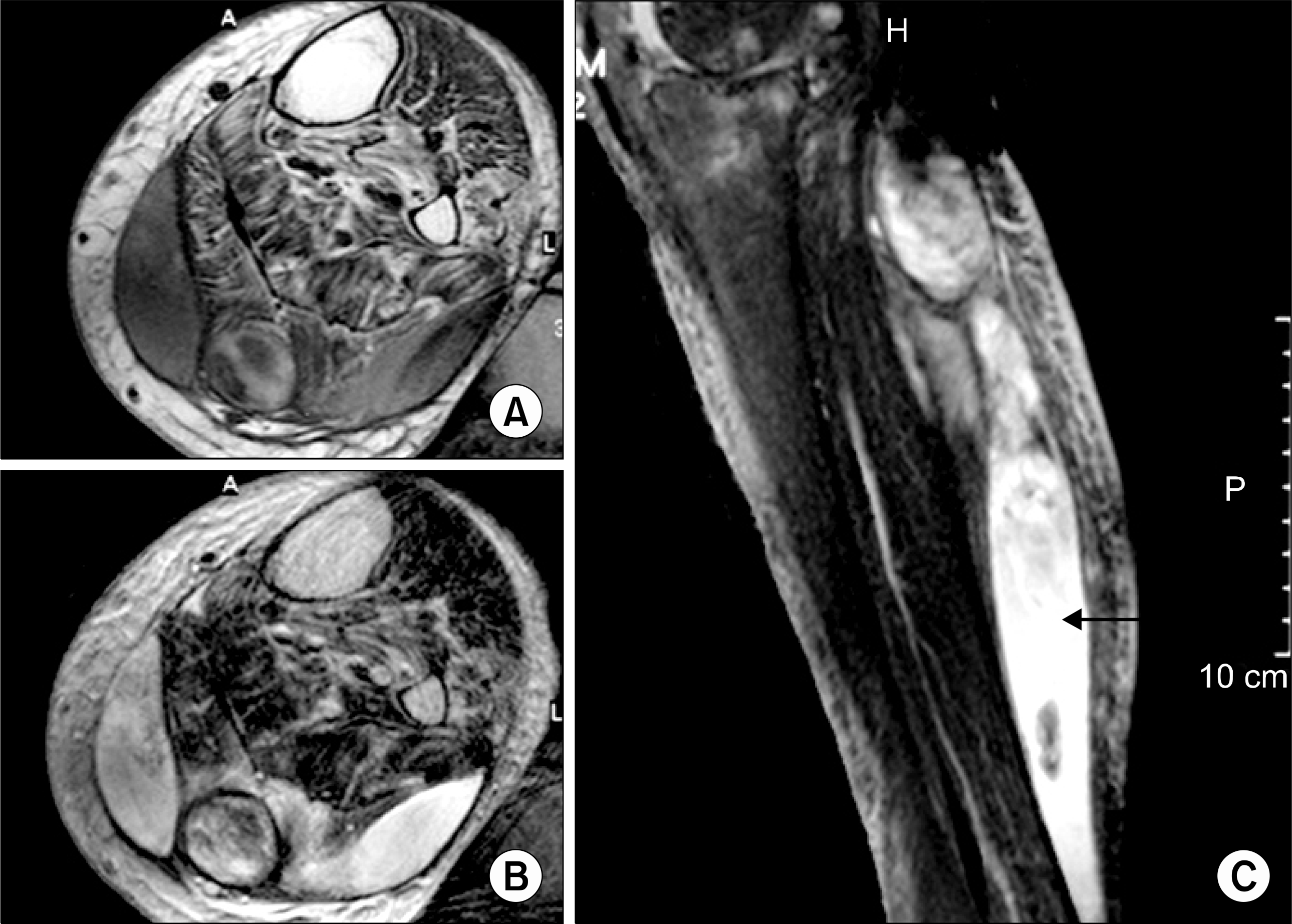
Table 1.
Causes of calf symptoms
Table 2.
Characteristics of the complicated popliteal cysts
 XML Download