

 PDF
PDF ePub
ePub Citation
 Print
Print


Abstract
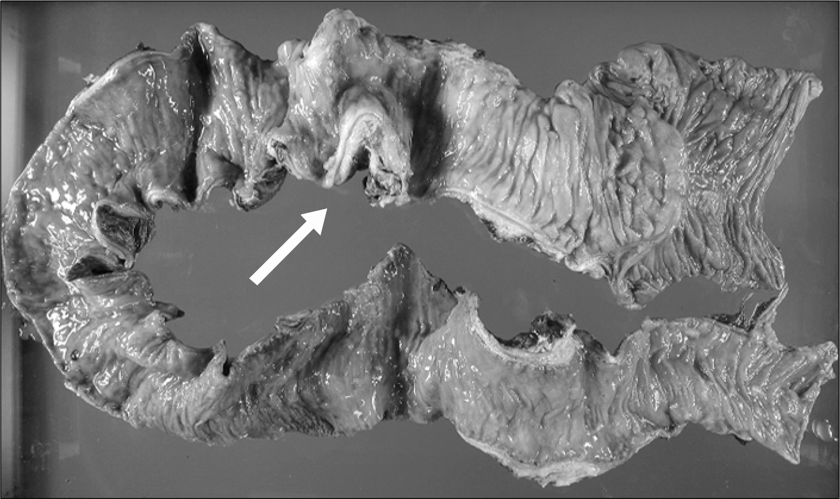
Idiopathic sclerosing peritonitis is a rare disease characterized by fibrosis and adhesion of the peritoneum to loops of the small intestine. It may be the cause of an unusual surgical emergency such as small bowel ileus. It is diagnosed predominantly in female adolescents. We report the case of an idiopathic sclerosing peritonitis in Korea. A 38-year-old man visited emergency room for recurrent small bowel ileus and migrating mass like lesion. Computed tomography (CT) of abdomen showed acute peritonitis with a diffuse wall thickening of terminal ileum and extraluminal fluid collectionaround the terminal ileum. He underwent laparotomy. The ileocolectomy with adhesiolysis was performed and its pathological examination revealed the characteristic findings of idiopathic sclerosing peritonitis. Symptoms recurred 2 months after surgery, but improved with steroid treatment.
References
1. Foo KT, Ng KC, Rauff A, Foong WC, Sinniah R. Unusual small intestinal obstruction in adolescent girls: the abdominal cocoon. Br J Surg. 1978; 65:427–30.
2. McLaughlin K, Butt G, Madi A, McMillan M, Mactier R. Sclerosing peritonitis occurring in a hemodialysis patients. Am J Kidney Dis. 1996; 27:729–32.
3. Burstein M, Galun E, Ben-Chetrit E. Idiopathic sclerosing peritonitis in a man. J Clin Gastroenterol. 1990; 12:698–701.

4. Narayanan R, Kabra SG, Bhargava BN, Sangal BC. Idiopathic sclerosing encapsulating peritonitis. Lancet. 1989; 2:127–9.
5. Kobayashi TK, Ueda M, Nishino T, Nishida S, Yeneyama C, Watanabe S. Appearance of ‘collagen balls' in ascitic fluid cytology with abdominal cocoon (encapsulating peritonitis) [letter]. Diagn Cytopathol. 1997; 16:469–70.
6. Suh WN, Lee SK, Chang H, Hwang HJ, Hyung WJ, Park YN, et al. Sclerosing encapsulating peritonitis (abdominal cocoon) after abdominal hysterectomy. Korean J Intern Med. 2007; 22:125–9.
7. Marshall AJ, Baddeley H, Barritt DW, Davies JD, Lee RE, Low-Beer TS, et al. Practolol peritonitis. A study of 16 cases and a survey of small bowel function in patients taking beta adrenergic blockers. Q J Med. 1977; 181:135–49.
8. Marigold J, Pounder RE, Pemberton J, Thompson RPH. Propranolol, Oxprenolol and sclerosing peritonitis. Br Med J. 1982; 284:870.
9. Afthentopoulos IE, Passadakis P, Oreopoulos DG, Bargman J. Sclerosing peritonitis in continuous ambulatory peritoneal dialysis patients: one center's experience and review of the literature. Adv Ren Repalce Ther. 1998; 5:165–7.
10. Holland P. Sclerosing encapsulating peritonitis in chronic ambulatory peritoneal dialysis. Clin Radiol. 1990; 41:19–23.
11. Kaushik R, Punia R, Mohan H, Attri AK. Tuberculous abdominal cocoon-a report of 6 cases and review of the literature. World J Emerg Surg. 2006; 1:18.
12. Pepels MJ, Peters FP, Mebis JJ, Ceelen TL, Hoofwijk AG, Erdkamp FL. Sclerosing peritonitis: an unusual cause of ascites in a patient with systemic lupus erythematosus. Neth J Med. 2006; 64:346–9.
13. Masuda C, Fujii Y, Kamiya T, Miyamoto M, Nakahara K, Hattori S, et al. Idiopathic sclerosing peritonitis in a man. Intern Med. 1993; 32:552–5.
14. Xu P, Chen LH, Li YM. Idiopathic sclerosing encapsulating peritonitis (or abdominal cocoon): a report of 5 cases. World J Gastroenterol. 2007; 13:3649–51.
15. Cleffken B, Sie G, Riedl R, Heineman E. Idiopathic sclerosing encapsulating peritonitis in a young female-diagnosis of abdominal cocoon. J Pediatr Surg. 2008; 43:27–30.
Fig. 1.
Simple abdomen erect and supine views show severe small bowel dilatation, suggesting small bowel obstruction.
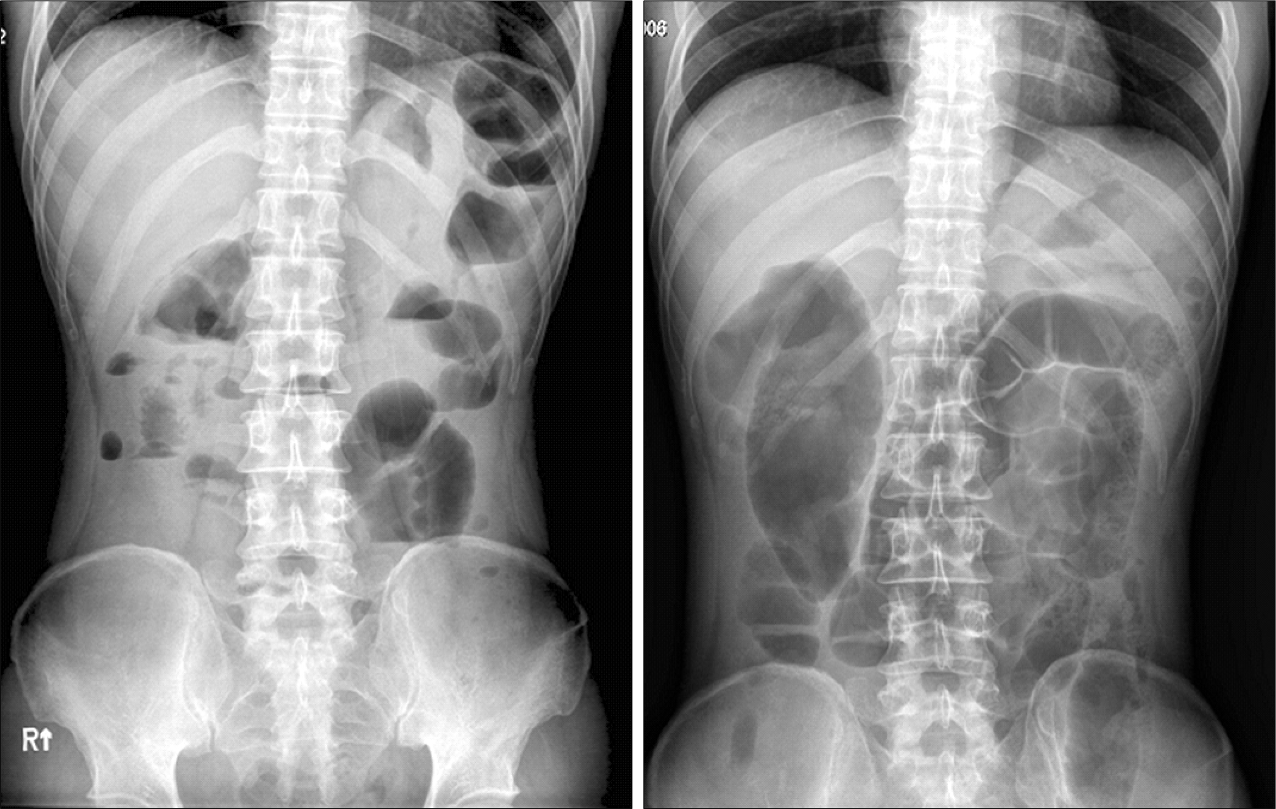
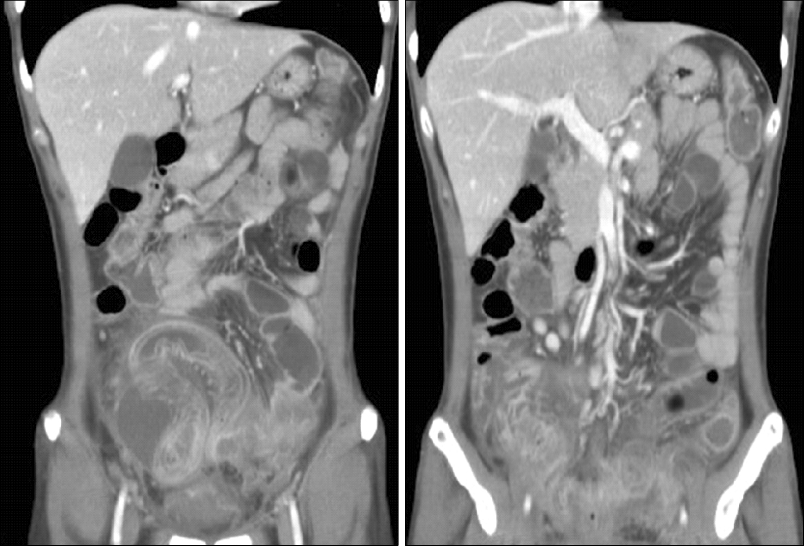
Fig. 2.
Abdominal CT. The terminal ileum shows diffuse wall thickening. It reveals the lower abdominal peritonitis and extraluminal fluid collection around the terminal ileum.

 XML Download