Abstract
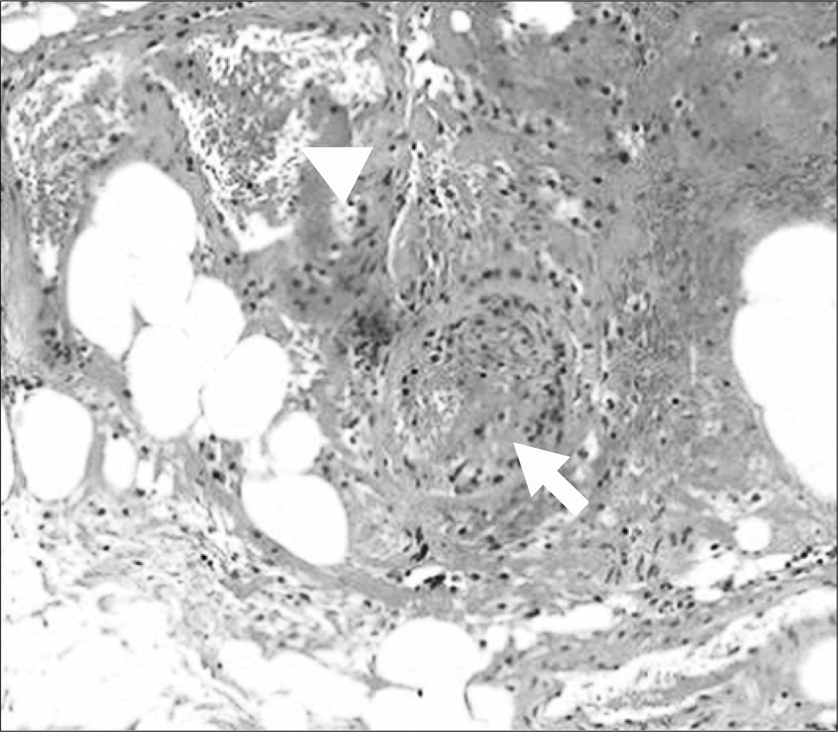
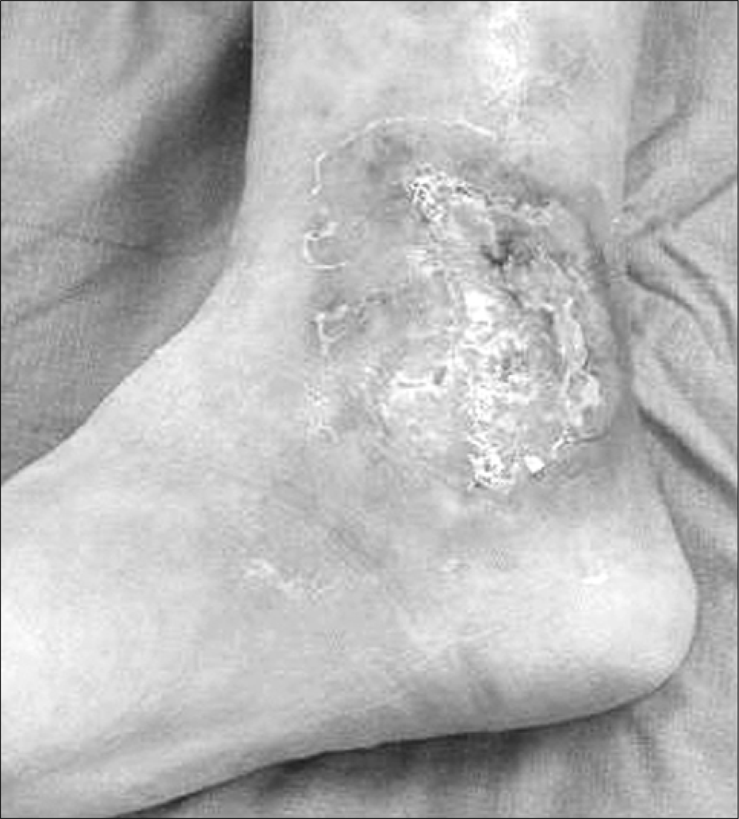
Antiphospholipid syndrome (APS) is characterized by vascular thrombosis in association with elevated titers of antiphospholipid antibodies. Leg ulcers are a considered to be a cutaneous manifestation of APS due to thrombosis of small to medium sized vessels. We report a case of necrotic non-healing, ankle ulcers mimicking pyoderma gangrenosum associated with APS in 50-year-old man. He had a past history of autoimmune thrombocytopenia and cerebral infarction. Laboratory findings showed a circulating lupus anticoagulant, positive anticardiolipin antibodies as well as anti-dsDNA and anti-Sm antibodies. Skin biopsy of ulcer lesions showed thrombotic vasculopathy of medium sized vessels with minimal leukocyte infiltration. Ulcers were successfully treated with surgical debridement and subsequent skin graft along with anticoagulation therapy.
References
1. Wilson WA, Gharavi AE, Koike T, Lockshin MD, Branch DW, Piette JC, et al. International consensus statement on preliminary classification criteria for definite antiphospholipid syndrome. Report of International workshop. Arthritis Rheum. 1999; 42:1309–11.
2. Diogenes MJ, Diogenes PC, de Morias Carneiro RM, Neto CC, Duarte FB, Holanda RR. Cutaneous manifestation associated with antiphospholipid antibodies. Int J Dermatol. 2004; 43:632–7.
3. Gibson GE, Su WPS, Pittelkow MR. Antiphospholipid syndrome and the skin. J Am Acad Dermatol. 1997; 36:970–82.

4. Alegre VA, Gastineau DA, Winkelmann RK. Skin lesions associated with circulating lupus anticoagulant. Br J Dermatol. 1989; 120:419–29.

5. Zamiri M, Griffiths D, Jarrett P. Recalcitrant leg ulcer as the initial manifestation of antiphospholipid syndrome in a 14-year-old boy. Intern Med J. 2001; 31:315–6.

6. Freedman AM, Phelps RG, Lebwohl M. Pyoderma gangrenosum associated with anticardiolipin antibodies in a pregnant patient. Int J Dermatol. 1997; 36:205–12.

7. Babe KS Jr, Gross AS, Leyva WH, King LE Jr. Pyoderma gangrenoso associated with antiphospholipid antibodies. Int J Dermatol. 1992; 31:588–90.
8. Cardinali C, Caproni M, Bernacchi E, Amato L, Fabbri P. The spectrum of cutaneous manifestations of lupus erythematosus-The Italian experience. Lupus. 2000; 9:417–23.
9. Kapadia N, Haroon TS. Cutaneous manifestation of systemic lupus erythematosus: Study from Lahore, Pakistan. Int J Dermatol. 1996; 35:408–9.
10. Alarcon-Segovia D, Deleze M, Oria CV, Sanchez-Guerrero J, Gomez-Pacheco L, Cabiedes J, et al. Antiphospholipid antibodies and the antiphospholipid syndrome in systemic lupus erythematosus. A prospective analysis of 500 consecutive patients. Medicine (Baltimore). 1989; 68:353–65.
11. Alarcon-Segovia D, Perez-Vazquez ME, Villa AR, Drenkard C, Cabiedes J. Preliminary classification criteria for the antiphospholipid syndrome within systemic lupus erythematosus. Semin Arthritis Rheum. 1992; 21:275–86.
12. Stephansson EA, Niemi KM, Jouhikainen T, Vaarala O, Palosuo T. Lupus anticoagulant and the skin. A longterm follow-up study of SLE patients with special reference to histopathological findings. Acta Derm Venereol. 1991; 71:416–22.
13. 김범경, 최상태, 손명균, 이광훈, 이상원, 정세진 등. 전신홍반루푸스에 의한 속발성 항인지질증후 군에 동반된 정맥성 족부궤양 치험 1예. 대한류마 티스학회지. 2007; 14:71–7.
14. Tishler M, Papo J, Yaron M. Skin ulcer as the presenting symptom of primary antiphospholipid syndrome-resolution with anticoagulant therapy. Clin Rheumatol. 1995; 14:112–4.

15. Gertner E, Lie JT. Systemic therapy with fibrinolytic agents and heparin for recalcitrant nonhealing cutaneous ulcer in the antiphospholipid syndrome. J Rheumatol. 1995; 21:2159–61.
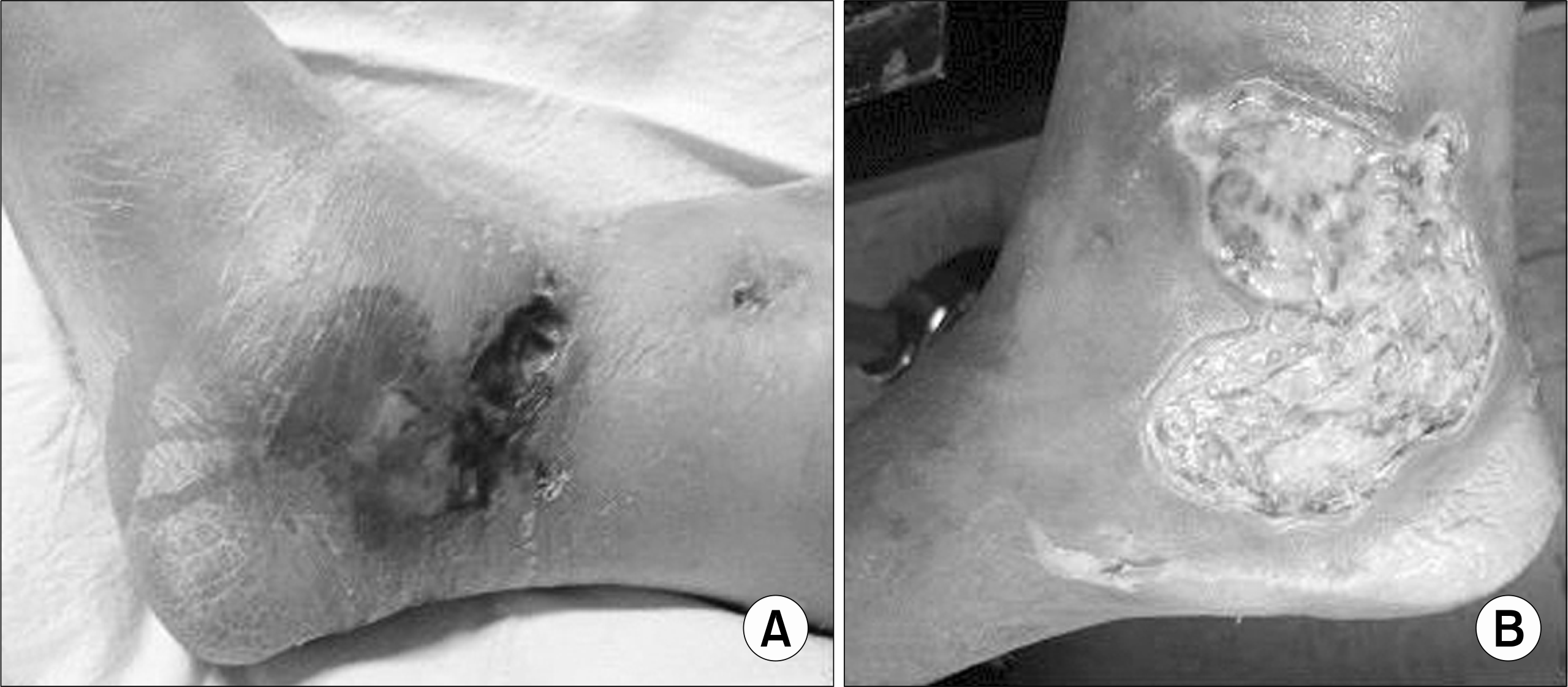
Fig. 1.
Skin ulceration over the right medial malleolus. Multiple ulcers with violaceous overhanging borders and a necrotic edge (A). Fibrinous exudate and granulation tissue on ulcer base after surgical debridement (B).


 PDF
PDF ePub
ePub Citation
 Print
Print


 XML Download