Abstract
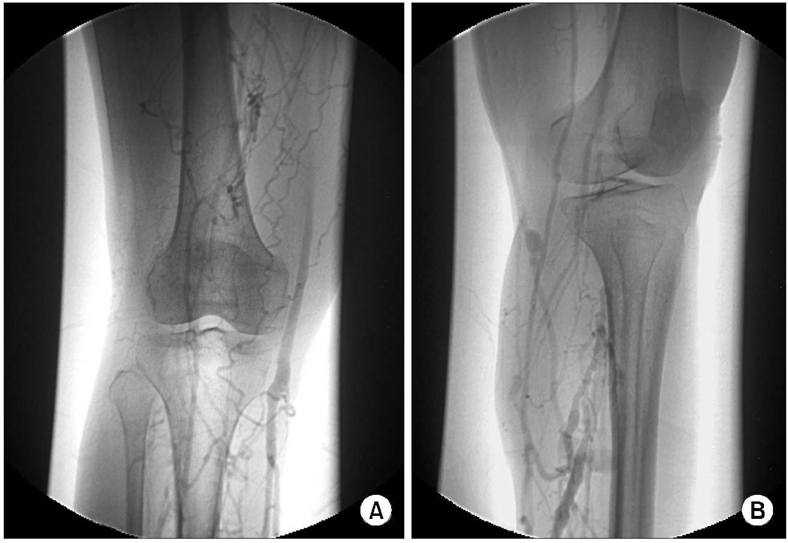
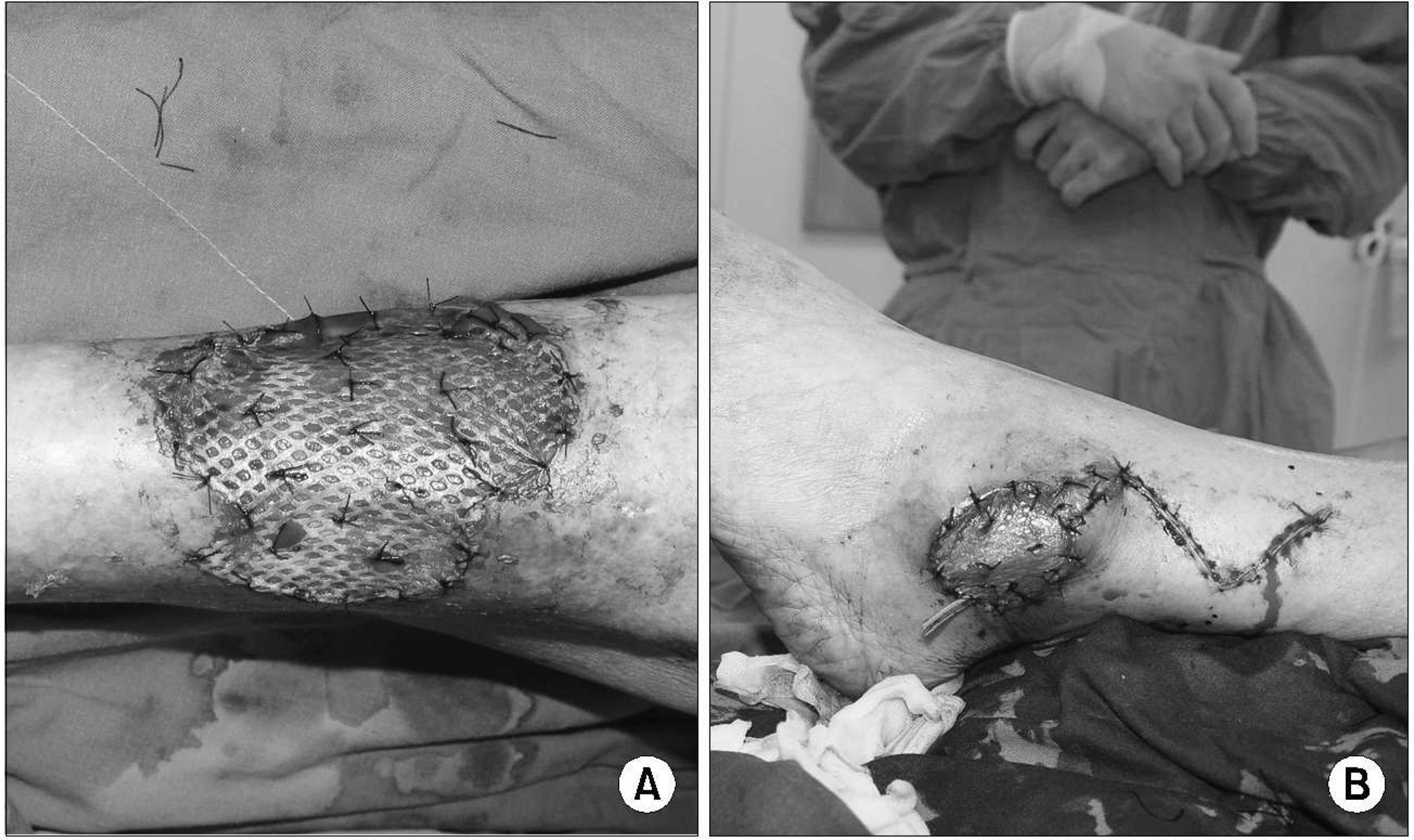
Antiphospholipid syndrome is an autoimmune disorder characterized by recurrent arterial or venous thrombosis, and pregnancy loss. A 57-year-old woman was admitted for aggravation of both leg ulcers. Venogram showed chronic venous obstructions at both lower extremities, and chest x-ray and computed tomography revealed serositis in pericardium and pleura. The laboratory tests revealed pancytopenia, and positive tests for antinuclear antibody, anti-dsDNA antibody, lupus anticoagulant and anticardiolipin antibody, which led to a diagnosis of anti- phospholipid syndrome secondary to systemic lupus erythematous. After medical treatments by anticoagulation and immunosuppression, and surgical managements including subtotal skin graft and local flap surgery, leg ulcers had been successfully treated without recurrence. Recognition of antiphospholipid syndrome as a cause of venous ulcer and the treatment plans including anticoagulation and surgical management is important in proper managements.
REFERENCES
Hughes GR., Harris NN., Gharavi AE. The anticardiolipin syndrome. J Rheumatol. 1986. 13:486–9.
Lim W., Crowther MA., Eikelboom JW. Management of antiphospholipid antibody syndrome: a systematic review. JAMA. 2006. 295:1050–7.
Love PE., Santoro SA. Antiphospholipid antibodies: anticardiolipin and the lupus anticoagulant in systemic lupus erythematosus (SLE) and in non-SLE disorders. Prevalence and clinical significance. Ann Intern Med. 1990. 112:682–98.
Trent JT., Falabella A., Eaglstein WH., Kirsner RS. Venous ulcers: pathophysiology and treatment options. Ostomy Wound Manage. 2005. 51:38–54.
Omar AA., Mavor AID., Jones AM., Homer-Vanniasinkam S. Treatment of Venous Leg Ulcers with Dermagraft (R). Eur J Vasc Endovasc Surg. 2004. 27:666–72.
Patel NP., Labropoulos N., Pappas PJ. Current management of venous ulceration. Plast Reconstr Surg. 2006. 117:254–60.

Bitsch M., Saunte DM., Lohmann M., Holstein PE., Jorgensen B., Gottrup F. Standardised method of surgical treatment of chronic leg ulcers. Scand J Plast Reconstr Surg Hand Surg. 2005. 39:162–9.

Lim JS., Kim HJ., Joo HS., Choi YS. Treatment of Chronic Wound in a Patient with Systemic Vasculitis. J Korean Soc Plast Reconstr Surg. 2006. 33:116–9.
Khamashta MA., Cuadrado MJ., Mujic F., Taub NA., Hunt BJ., Hughes GR. The management of thrombosis in the antiphospholipid-antibody syndrome. N Engl J Med. 1995. 332:993–7.

DeMarco P., Singh I., Weinstein A. Management of the antiphospholipid syndrome. Curr Rheumatol Rep. 2006. 8:114–20.

McClain MT., Arbuckle MR., Heinlen LD., Dennis GJ., Roebuck J., Rubertone MV, et al. The prevalence, onset, and clinical significance of antiphospholipid antibodies prior to diagnosis of systemic lupus erythematosus. Arthritis Rheum. 2004. 50:1226–32.

Erkan D., Lockshin MD. New treatments for antiphospholipid syndrome. Rheum Dis Clin North Am. 2006. 32:129–48.

Kirsner RS., Mata SM., Falanga V., Kerdel FA. Split-thickness skin grafting of leg ulcers. The University of Miami Department of Dermatology's experience (1990-1993). Dermatol Surg. 1995. 21:701–3.

15). Douglas WS., Simpson NB. Guidelines for the management of chronic venous leg ulceration. Report of a multidisciplinary workshop. British Association of Dermatologists and the Research Unit of the Royal College of Physicians. Br J Dermatol. 1995. 132:446–52.
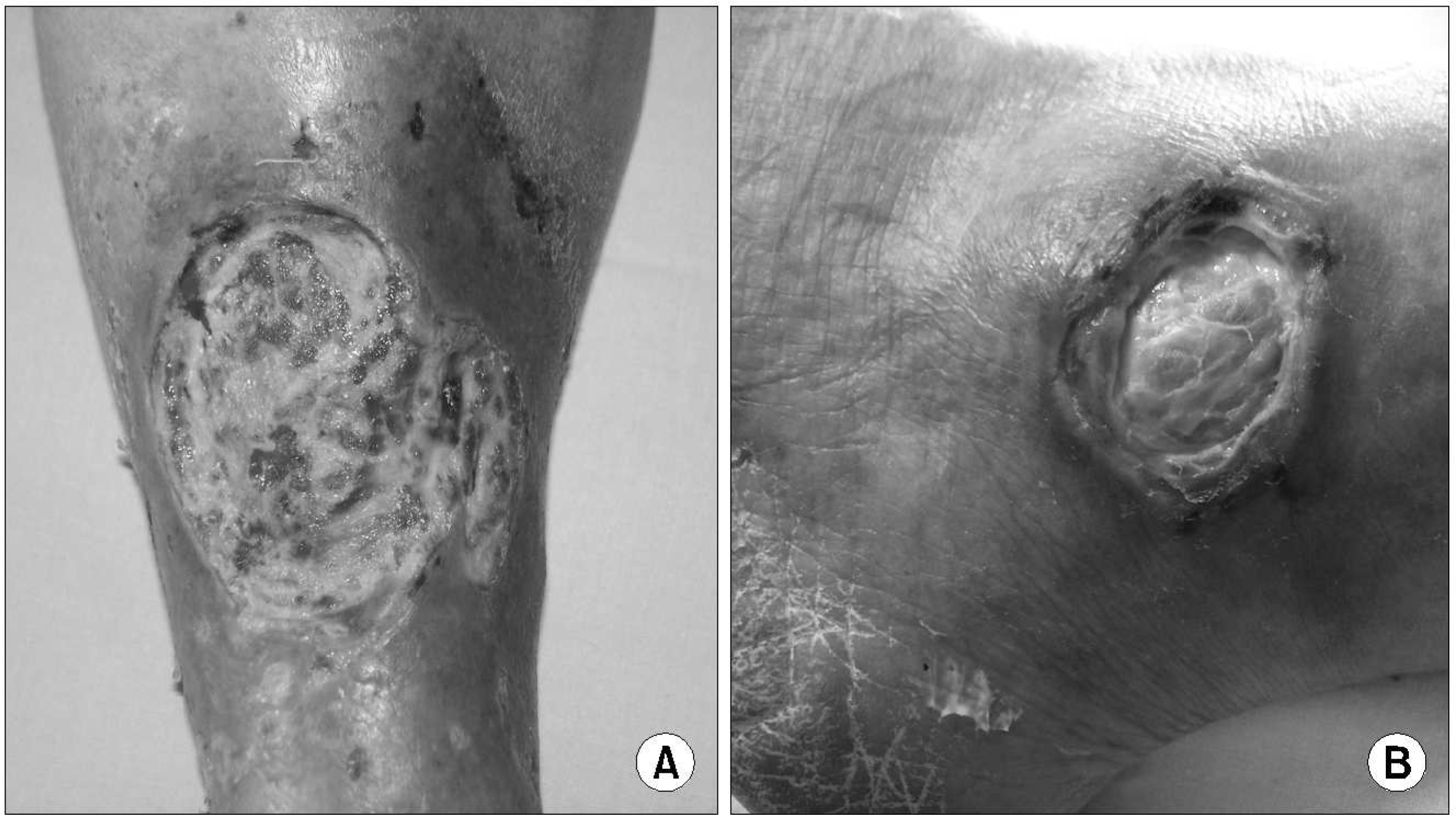
Fig. 1.
There was 3 X4 cm sized ulcer at right lower tibia (A), and 2 X3 cm sized ulcer at left malleus area (B).

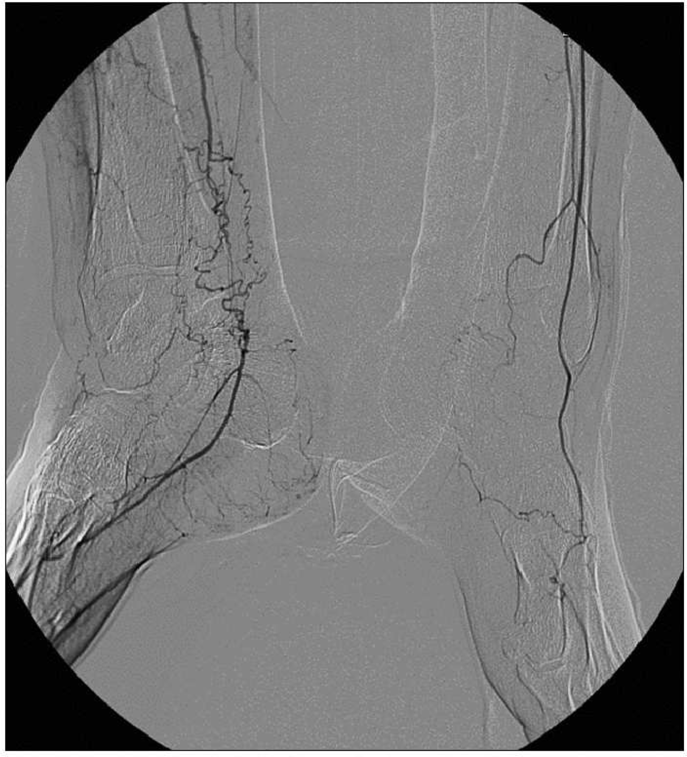
Fig. 2.
Distal obstructions of left posterior tibial artery, and right anterior · posterior tibial artery were found in the arteriogram.


 PDF
PDF ePub
ePub Citation
 Print
Print


 XML Download