

 PDF
PDF ePub
ePub Citation
 Print
Print


Abstract
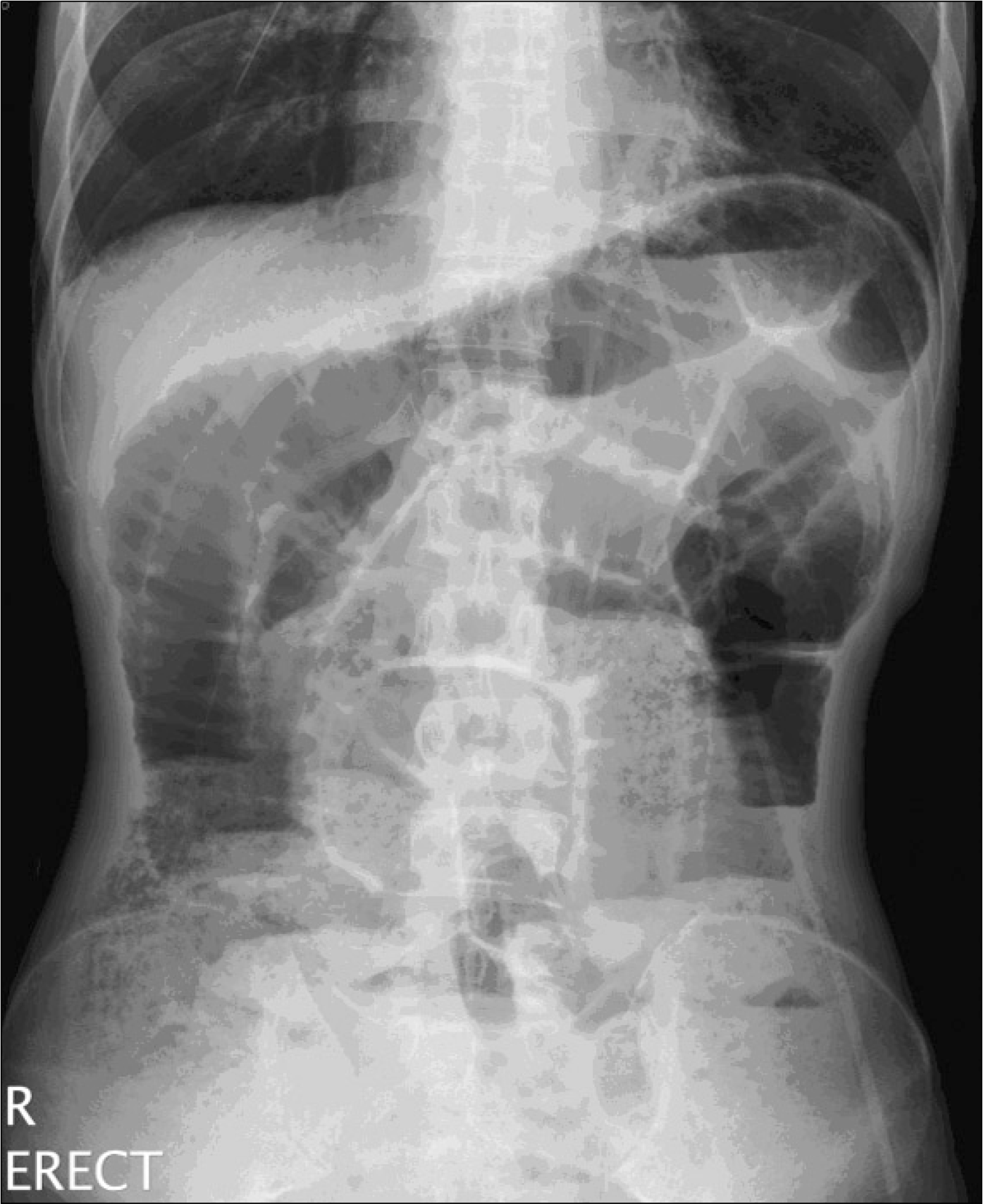
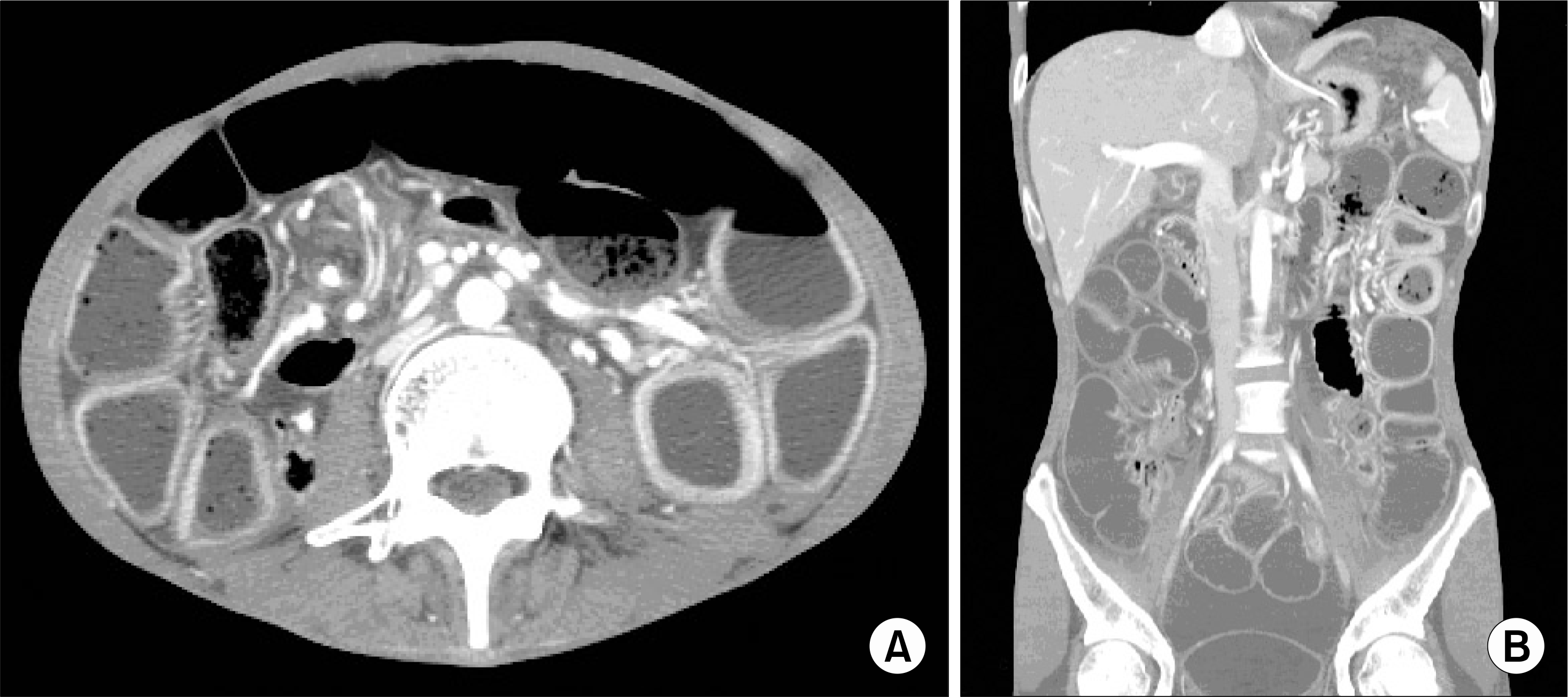
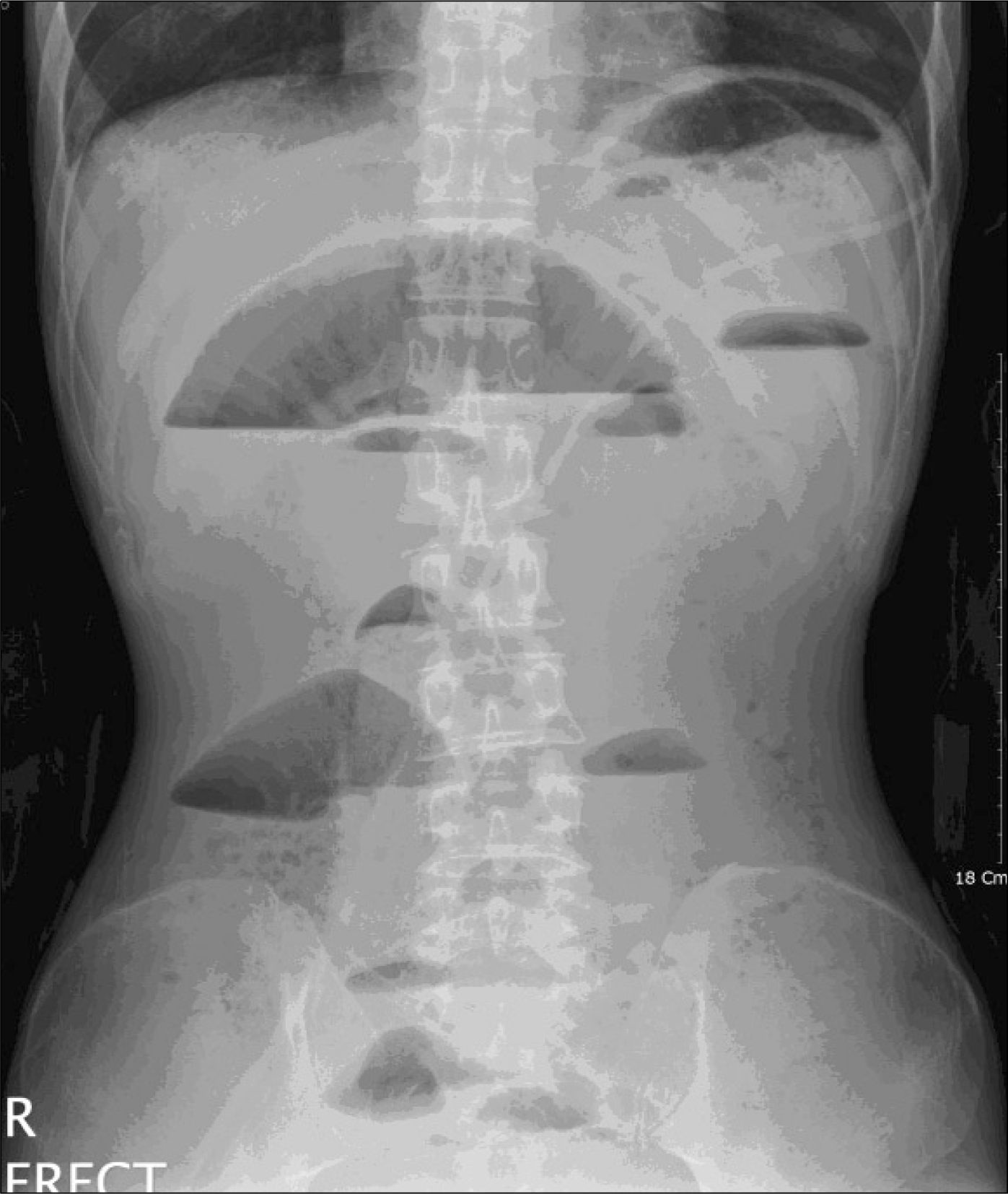
Systemic sclerosis is an autoimmune disease, characterized by inflammation, microangiopathy and fibrosis in the skin and various intestinal organs. Gastrointestinal involvement is one of the major causes of serious morbidity, and next to the skin, the gastrointestinal tract is the most commonly involved organ. While esophageal involvement is most common gastrointestinal manifestation, the involvement of the small intestine and colon is less common but may lead to life-threatening complications like chronic pesudoobstruction or pneumatosis cystoids intestinalis. Here, we describe a case of progressive systemic sclerosis associated with intestinal pseudoobstruction. 28 year-old male presented abdominal pain and vomiting and he was diagnosed as having intestinal pseudoobstruction. His symptoms were well managed using the combination of octreotide, a long-acting somatostatin analogue, and erythromycin.
REFERENCES
1). LeRoy EC., Black C., Fleischmajer R., Jablonska S., Kreig T., Medsger TA Jr, et al. Scleroderma (systemic sclerosis): classification, subsets and pathogenesis. J Rheumatol. 1988. 15:202–5.
2). Clements P., Lachenbruch P., Seibold J., White B., Weiner S., Martin R, et al. Inter- and intraobserver variability of the total thickness score (modified Rodnan TSS) in systemic sclerosis. J Rheumatol. 1995. 22:1281–5.
3). Sallam H., McNearney TA., Chen JD. Systematic review: pathophysiology and management of gastrointestinal dysmotility in systemic sclerosis (scleroderma). Aliment Pharmacol Ther. 2006. 23:691–712.

4). Ponge T., Bruley des Varannes S. Digestive involvement of scleroderma. Rev Prat. 2002. 52:1896–900.
5). Mittag M., Haustein UF. Progressive systemic scleroderma-prognosis determining involvement of internal organ systems. Hautarzt. 1998. 49:545–51.
6). Folwaczny C., Voderholzer W., Riepl RL., Schindlbeck N. Clinical aspects, pathophysiology, diagnosis and therapy of gastrointestinal manifestations of progressive systemic scleroderma. Z Gastroenterol. 1996. 34:497–508.
7). Cohen S., Fisher R., Lipshutz W., Turner R., Myers A., Schumacher R. The pathogenesis of esophageal dysfunction in scleroderma and Raynaud's disease. J Clin Invest. 1972. 51:2663–8.
8). Quiroz ES., Flannery MT., Martinez EJ., Warner EA. Pneumatosis cystoides intestinalis in progressive systemic sclerosis: a case report and literature review. Am J Med Sci. 1995. 310:252–5.
9). Emmanuel AV., Shand AG., Kamm MA. Erythromycin for the treatment of chronic intestinal pseudo-obstruction: description of six cases with a positive response. Aliment Pharmacol Ther. 2004. 19:687–94.
10). Itou H., Iizuka M., Shindo K., Konno S., Watanabe S. A case of pseudoobstruction of the intestine associated with scleroderma dramatically responding to antibiotics. J Gastroenterol. 2006. 41:711–2.
11). Harris AG. Somatostatin and somatostatin analogues: pharmacokinetics and pharmacodynamic effects. Gut. 1994. 35((3 Suppl):):S1–4.
12). Kobayashi T., Kobayashi M., Naka M., Nakajima K., Momose A., Toi M. Response to octreotide of intestinal pseudoobstruction and pneumatosis cystoides intestinalis associated with progressive systemic sclerosis. Intern Med. 1993. 32:607–9.
13). Perlemuter G., Cacoub P., Chaussade S., Wechsler B., Couturier D., Piette JC. Octreotide treatment of chronic intestinal pseudoobstruction secondary to connective tissue diseases. Arthritis Rheum. 1999. 42:1545–9.
14). Verne GN., Eaker EY., Hardy E., Sninsky CA. Effect of octreotide and erythromycin on idiopathic and scleroderma-associated intestinal pseudoobstruction. Dig Dis Sci. 1995. 40:1892–901.
15). 김채기: 류헌모: 권중구: 이창형: 송용호: 최정윤. Systemic sclerosis sine sclerderma 1예. 대한류마티스학회지. 2000. 7:313–8.
 XML Download