

 PDF
PDF ePub
ePub Citation
 Print
Print


Abstract
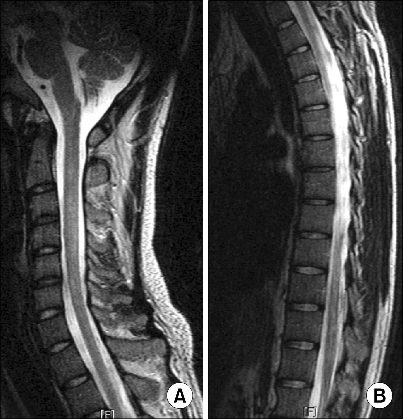
Neuromyelitis optica (NMO) is an idiopathic inflammatory demyelinating disease, characterized by optic neuritis and myelitis. NMO is a very uncommon and serious neurologic manifestation of systemic lupus erythematous (SLE). We report a 28-year-old man with NMO as neuropsychiatry manifestation of SLE. He was diagnosed as lupus nephritis at 16-year-old. He had optic neuritis at three years and seven months ago. Oral prednisolone was tapered off according to the improved eye symptoms. Two months later, he visited rheumatology clinics for urinary disturbance and paresthesia on both feet. A spinal magnetic resonance imaging revealed increased signal intensity in T2-weighted images from second to sixth cervical level and from eleventh to twelfth thoracic level. We diagnosed neuromyelitis optica and treated with intravenous cyclophosphamide therapy monthly for three times. He was discharged without any neurological deficits and has been followed up.
REFERENCES
1). Handly JG. Neuropsychiatric lupus. Rheum Dis Clin N Am. 2005. 31:273–98.
2). Kovacs B., Lafferty TL., Brent LH., DeHoratius RJ. Transverse myelopathy in systemic lupus erythematosus: an analysis of 14 cases and review of the literature. Ann Rheum Dis. 2000. 59:120–4.

3). Mandler RN. Neuromyelitis optica-Devic's syndrome, update. Autoimmun Rev. 2006. 5:537–43.
4). 안태범: 윤병우. 반복적인척수염과시신경염을보인전신성홍반성낭창1예. 대한신경과학회지. 2000. 18:65760.
5). 정상준: 박기형: 김희태: 김명호. 폐결핵이후발생한시속신경척수염1 예. 대한신경과학회지. 2000. 18:85–8.
6). Wingerchuk DM., Lennon VA., Pittock SJ., Lucchinetti CF., Weinshenker BG. Revised diagnostic criteria for neuromyelitis optica. Neurology. 2006. 66:1485–9.
7). Minagar A., Alexander JS., Fowler MR., Long AC., Kelley RE. Devic disease: clinical course, pathophysiology, and management. Pathophysiology. 2002. 9:33–40.
8). Lucchinetti CF., Mandler RN., McGavern D., Bruck W., Gleich G., Ransohoff RM, et al. A role for humoral mechanisms in the pathogenesis of Devic's neuromyelitis optica. Brain. 2002. 125:1450–61.
9). Mok CC., Lau CS., Chan EY., Wong RW. Acute transverse myelopathy in systemic lupus erythematosus: clinical presentation, treatment, and outcome. J Rheumatol. 1998. 25:467–73.
10). Hagiwara N., Toyoda K., Uwatoko T., Yasumori K., Ibayashi S., ᄋkada Y. Successful high dose glucocorticoid treatment for subacute neuromyelitis optica with systemic lupus erythematosus. Intern Med. 2005. 44:998–1001.
11). Keegan M., Pineda AA., McClelland RL., Darby CH., Rodriguez M., Weinshenker BG. Plasma exchange for severe attacks of CNS demyelination: predictors of response. Neurology. 2002. 58:143–6.
12). Mandler RN., Ahmed W., Dencoff JE. Devic's neuromyelitis optica: a prospective study of seven patients treated with prednisone and azathioprine. Neurology. 1998. 51:1219–20.
13). Barile-Fabris L., Ariza-Andraca R., 이guin-ᄋrtega L., Jara LJ., Fraga-Mouret A., Miranda-Limon JM, et al. Controlled clinical trial of IV cyclophosphamide versus IV methylprednisolone in severe neurological manifestations in systemic lupus erythematosus. Ann Rheum Dis. 2005. 64:620–5.
14). Lennon VA., Wingerchuk DM., Kryzer TJ., Pittock SJ., Lucchinetti CF., Fujihara K, et al. A serum autoantibody marker of neuromyelitis optica: distinction from multiple sclerosis. Lancet. 2004. 364:2106–12.
15). Protti A., Erminio C., Piccolo I., Spreafico C., Colombo F., Ghezzi A. An unusual case with relapsing neuromyelitis optica associated with undifferentiated connective tissue disease. Neurol Sci. 2004. 25(suppl 4):S383–5.
Fig. 1.
(A) Fundus photography as the frist episode of optic neuritis. Left fundus photography shows the hyperemia, edematous change and blurring around optic disc and right fundus photography shows normal optic disc. (B) Fundus photography as the second episode of optic neuritis. Right fundus photography shows the hyperemia around optic disc and left fundus photography shows the pale optic disc.

 XML Download