

 PDF
PDF ePub
ePub Citation
 Print
Print


Abstract
Here we describe a male patient who attained normal height despite combined hypopituitarism with an abnormal growth hormone-insulin-like growth factor (IGF)-I axis. When he was an 18-year-old, he presented with a short stature and underdeveloped external genitalia. The patient had not undergone normal pubertal development and he displayed a height below the fifth percentile. Hormonal and radiological studies revealed the findings of severe anterior pituitary hormone deficiency and an atrophic pituitary gland. There had been no recent follow-ups with the patient or medical treatment since that time. In the current presentation, the patient, now 22 years of age, had attained normal height, yet he remained prepubertal and showed manifestations of delayed bone age and combined hypopituitarism. In addition, the patient's IGF-II levels were increased for his age.
References
1. Low MJ. Neuroendocrinology. In: Kronenbeg HM, Melmed S, Polonsky KS, Larsen PR ed. Williams textbook of endocrinology. 11th ed. pp.85–154. Philadelphia, WB Sounders Co.,. 2008.
2. Kenny FM, Guyda HJ, Wright JC, Friesen HG. Prolactin and somatomedin in hypopituitary patients with “catch up” growth following operations for craniopharyngioma. J Clin Endocrinol Metab. 36:378–380. 1973.
3. Blethen SL, Weldon VV. Outcome in children with normal growth following removal of a craniopharyngioma. Am J Med Sci. 292:21–24. 1986.

4. Tiulpakov AN, Mazerkina NA, Brook CG, Hindmarsh PC, Peterkova VA, Gorelyshev SK. Growth in children with craniopharyngioma following surgery. Clin Endocrinol (Oxf). 49:733–738. 1998.
5. Bucher H, Zapf J, Torresani T, Prader A, Froesch ER, Illig R. Insulin-like growth factors I and II, prolactin, and insulin in 19 growth hormone-deficient children with excessive, normal, or decreased longitudinal growth after operation for craniopharyngioma. N Engl J Med. 309:1142–1146. 1983.
6. Bistritzer T, Chalew SA, Lovchik JC, Kowarski AA. Growth without growth hormone: the “invisible” GH syndrome. Lancet. 1:321–323. 1988.
7. Wada S, Minagawa A, Imamaki K, Suda S, Yamanaka K, Iitaka M, Katayama S. A patient of hypogonadotropic hypogonadism accompanied by growth hormone deficiency and decreased bone mineral density who attained normal growth. Intern Med. 39:641–645. 2000.
8. Lazar L, Dan S, Phillip M. Growth without growth hormone: growth pattern and final height of five patients with idiopathic combined pituitary hormone deficiency. Clin Endocrinol (Oxf). 59:82–88. 2003.
9. Papastathopoulou L, Tzanela M, Vlassopoulou V, Vassiliadi D, Thalassinos N. Untreated hypopituitarism due to absence of the pituitary stalk with normal adult height: report of two cases. Endocrine. 29:175–179. 2006.
10. Pavlou M, Tsatsoulis A, Efstathiadou Z, Bitsis S, Papadopoulou ZL. A study of the growth-promoting and metabolic effects of growth hormone (GH) in a patient with the “growth without GH” syndrome. Growth Horm IGF Res. 11:225–230. 2001.
11. Geffner ME, Lippe BM, Bersch N, Van Herle A, Kaplan SA, Elders MJ, Golde DW. Growth without growth hormone: evidence for a potent circulating human growth factor. Lancet. 1:343–347. 1986.
12. Murashita M, Tajima T, Nakae J, Shinohara N, Geffner ME, Fujieda K. Near-normal linear growth in the setting of markedly reduced growth hormone and IGF-1. A case report. Horm Res. 51:184–188. 1999.
13. Groop L, Segerlantz M, Bramnert M. Insulin sensitivity in adults with growth hormone deficiency and effect of growth hormone treatment. Horm Res. 64:45–50. 2005.
14. Lee PA, Witchel SF. The influence of estrogen on growth. Curr Opin Pediatr. 9:431–436. 1997.
15. Hill DJ, Milner RD. Insulin as a growth factor. Pediatr Res. 19:879–886. 1985.
16. Chao W, D'Amore PA. IGF2: epigenetic regulation and role in development and disease. Cytokine Growth Factor Rev. 19:111–120. 2008.
17. Constância M, Hemberger M, Hughes J, Dean W, Ferguson-Smith A, Fundele R, Stewart F, Kelsey G, Fowden A, Sibley C, Reik W. Placental-specific IGF-II is a major modulator of placental and fetal growth. Nature. 417:945–948. 2002.
18. Baker J, Liu JP, Robertson EJ, Efstratiadis A. Role of insulinlike growth factors in embryonic and postnatal growth. Cell. 75:73–82. 1993.
19. Wilson EM, Hsieh MM, Rotwein P. Autocrine growth factor signaling by insulinlike growth factor-II mediates MyoD-stimulated myocyte maturation. J Biol Chem. 278:41109–41113. 2003.
20. Minuto F, Palermo C, Arvigo M, Barreca AM. The IGF system and bone. J Endocrinol Invest. 28:8–10. 2005.
21. Assy N, Hochberg Z, Amit T, Shen-Orr Z, Enat R, Baruch Y. Growth hormone-stimulated insulinlike growth factor (IGF) I and IGF-binding protein-3 in liver cirrhosis. J Hepatol. 27:796–802. 1997.
22. Tomas FM, Knowles SE, Chandler CS, Francis GL, Owens PC, Ballard FJ. Anabolic effects of insulinlike growth factor-I (IGF-I) and an IGF-I variant in normal female rats. J Endocrinol. 137:413–421. 1993.
Fig. 1.
A photograph of the patient. Height was 176 cm and body weight was 72 kg (body mass index: 23.2 kg/m2). Waist/hip and upper/lower body ratios were 0.92 and 0.60, respectively. The patient presented with a micropenis, small scrotum, and no pubic hairs.
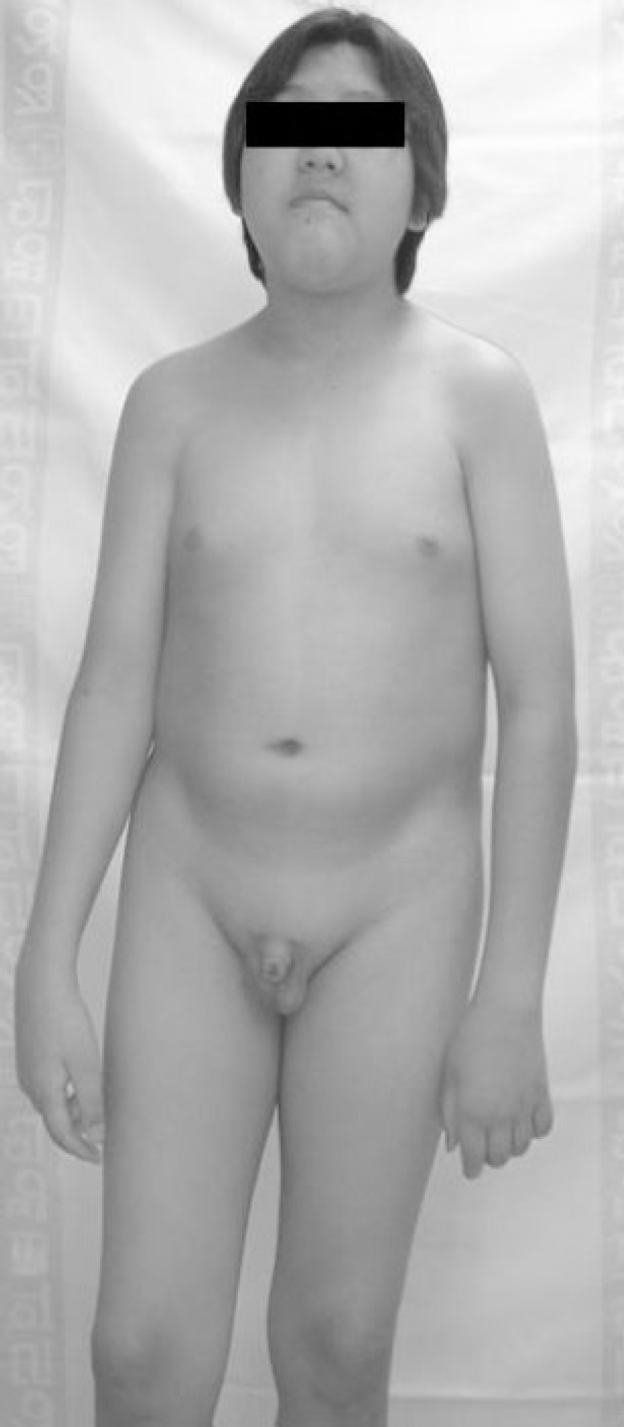
Fig. 2.
Patient growth curve. Until 16 years of age, the subject's height-for-age and weight-for-age remained below the 10th percentile. At age 18, the subject's height-for-age remained below the third percentile; however, the weight-for-age increased to the 25th percentile. At age 22, the patient gained mid-parental height and his weight-for-age increased to the 75th percentile. Closed squares represent height and closed circles represent weights, which were measured at 14, 15, 16, 17, 18 and 22 years of age.
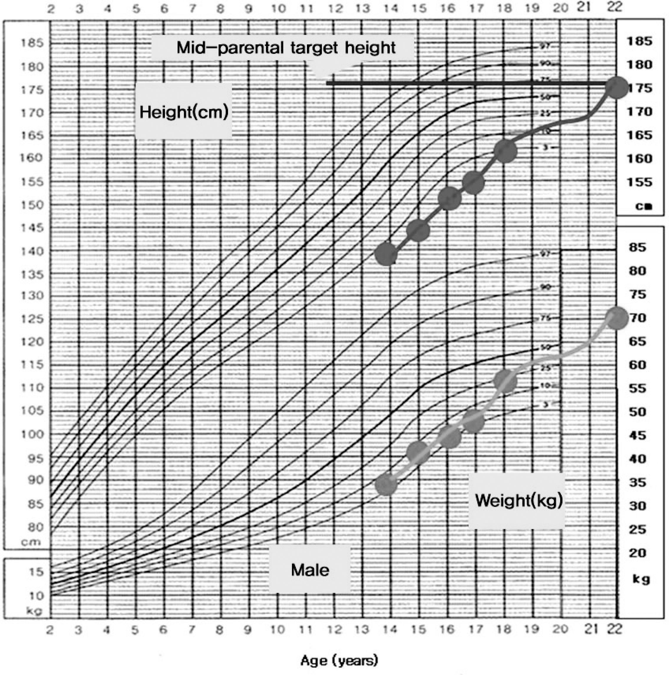
Fig. 3.
Sellar MRI findings. Sagittal view shows an interrupted or thin hypoplastic stalk (thick arrow) and an aplastic or hypoplastic hypophyseal gland (thin arrow).
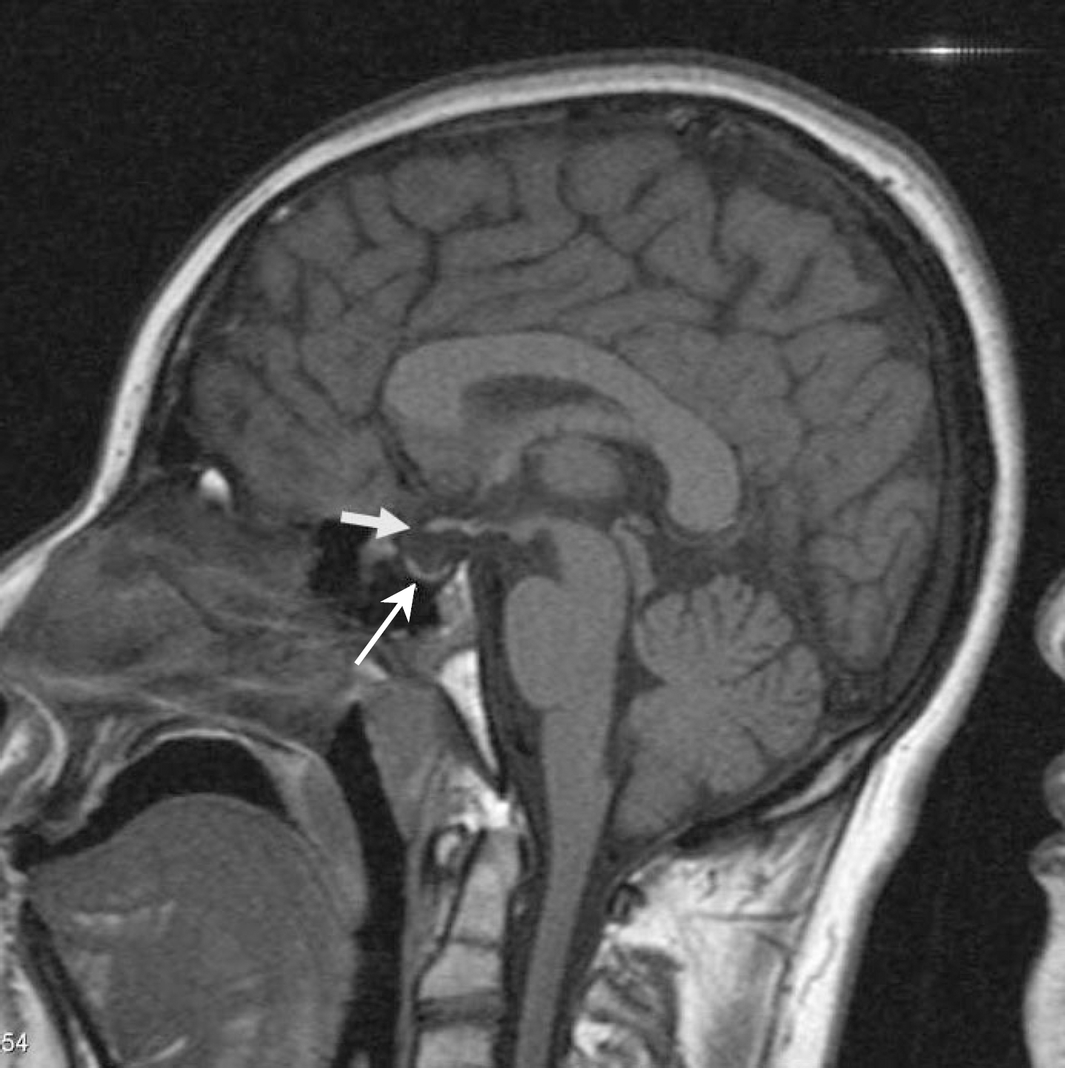
Table 1.
Combined pituitary stimulation tests performed during the first and second visits
 XML Download