

 PDF
PDF ePub
ePub Citation
 Print
Print


Introduction
Chorea is an involuntary movement disorder characterized by irregular, brief movements that flow from one body part to another in a non-stereotyped fashion. A spectrum of movements may be observed, including slower, distal, and writhing movements called athetosis. Although Huntington's Disease represents the prototypic choreic disorder[1], many other conditions including neurological, infectious, metabolic, endocrine, and autoimmune diseases may be accompanied by chorea[2~4]. Chorea secondary to hyperthyroidism is a rare manifestation, occurring in less than 2% of individuals with hyperthyroidism[5]. The neurological manifestations of Graves' disease include cognitive dysfunction and seizures, myasthenia gravis, thyrotoxic periodic paralysis, and peripheral neuropathy[6~10].
Recently, four cases of chorea secondary to Graves' disease have been reported. Each of the reported cases had different clinical manifestations and treatment modalities. Until now, the exact mechanism underlying this phenomenon remained unclear. We recently encountered a patient who initially presented with hyperthyroidism and subsequently developed progressive generalized chorea. The patient was diagnosed with chorea secondary to Graves' disease and improved following the initiation of steroid treatment.
Case
A 42-year-old female was admitted to the center complaining of continuous, involuntary movement in her left upper extremity and face that had been occurring for a month. She had a history of Graves' disease, which was diagnosed four years prior and treated with antithyroid medication. She had palpitations and weight loss, but she did not show any symptoms similar to chorea. Remission of the hyperthyroidism was achieved within two years. The patient discontinued the antithyroid medication but had her thyroid function checked regularly. Thyroid function tests were normal until four months prior to admission.
The patient's involuntary movements started a month prior to admission. Initially, the involuntary movement was very mild, and was only present in her face and distal left arm. The patient had also lost 5 kg of body weight over a single month prior to admission without concurrent lifestyle changes (i.e., diet and exercise). The choreic symptoms were aggravated 20 days prior to admission and during this time she visited two different private neurological clinics. Laboratory tests performed during the month before admission revealed undetectable levels of thyroid stimulating hormone (TSH) (< 0.01 µIU/mL), elevated serum T3 levels (654 ng/dL, normal range: 80~200 ng/dL) and elevated free T4 levels (8.30 ng/dL, normal range: 0.73~1.95 ng/dL). The patient was also positive for antimicrosomal antibodies (2961.2 U/mL, normal range: 0~60 U/mL) and TSH-Receptor antibodies (39.2 U/L, normal range: 0~1.5 U/L), but negative for antithyroglobulin antibodies (9.4 U/mL, normal range: 0~60 U/mL). Under the diagnosis of Graves' disease and chorea, she had been given anti-thyroid medication, anticholinergics, a dopamine agonist and antagonist, and a sedative agent. The patient's daily medications included propylthiouracil (PTU) (50 mg BID), propranolol HCl (40 mg BID), trihexyphenidyl HCl (2 mg TID), ropinirole (0.25 mg BID), clonazepam (0.5 mg BID), alprazolam (1 mg daily), and quetiapine fumarate (12.5 mg/25 mg daily). Although she had been taking these medications for 20 days, the patient's involuntary movement worsened. It was at this time that she visited our hospital.
On the day of admission, the patient's vital signs were stable except for a rapid heart rate of 92 beats per min. A diffuse goiter was noted and a neurological examination revealed the presence of involuntary movements of the face, tongue, and distal left arm. However, other neurological abnormalities, including dysphagia, were not observed. Laboratory tests demonstrated elevated T3 (273 ng/dL, normal range: 80~200 ng/dL), and free T4 (3.77 ng/dL, normal range: 0.73~1.95 ng/dL) levels, undetectable levels of TSH (< 0.01, normal range: 0.35~5.50 µIU/mL), a positive anti-microsomal antibody titer (> 3000 U/mL, normal range: 0~60 U/mL), a positive TBII of 51.16% (normal range: less than 10%), and a negative antithyroglobulin antibody titer of 8.20 U/mL.
Other autoimmune markers, including antinuclear antibodies, Anti-DNA antibodies, anticardiolipine antibodies, antiribonucleoprotein antibodies, Anti-Sjögren's Syndrome-A/Ro antibodies, Anti-Sjögren's Syndrome-B/La antibodies, p-antineutrophil cytoplasmic antibodies, c-antineutrophil cytoplasmic antibodies, and rheumatoid factor were all negative. Infection markers from blood and cerebrospinal fluid, including IgM anti-Cytomegalovirus antibodies, anti-Herpes simplex virus (HSV) antibodies, and IgM anti-Varicella zoster antibodies were also negative. Polymerase chain reaction (PCR) analyses from the same samples were also negative for mycobacterium tuberculosis complex and HSV type I. Therefore, we could exclude parainfectious autoimmune disorder and infectious chorea. On day 7 of admission, the 2- and 24-hour radioiodine thyroid uptake (RAIU) test values were 45.7% and 56.4%, respectively. Brain magnetic resonance imaging (MRI) with contrast enhancement was normal. Brain single-photon emission computed tomography (SPECT) did not show significantly increased uptake and an electroencephalography (EEG) was normal.
From day one of admission, the patient was infused intravenously with 1000 mg of Solu-medrol® for 5 days and given PTU (50 mg TID), propranolol HCl (40 mg TID), clonazepam (0.5 mg BID), and haloperidol (4.5 mg TID) daily. Choreic movement of the left side of the face, tongue, and distal left arm began to improve on the 4th day of Solu-medrol® infusion, and thyroid function tests were improved as compared with the lab tests taken on the day of admission. On the 5th day of Solu-medrol® infusion, the laboratory tests demonstrated normal T3 (132.64 ng/dL, normal range: 80~200 ng/mL) and elevated free T4 (3.98 ng/dL, normal range: 0.73~1.95 ng/dL) levels. TSH remained undetectable (< 0.01, normal levels: 0.4~3.1 µIU/mL) and anti-microsomal antibodies were elevated (1943.1 U/mL, normal range: 0~60 U/mL). After Solu-medrol® infusion, the patient was started on 60 mg of oral prednisolone acetate, daily. On the 7th day of steroid treatment, no choreic movement was observed and involuntary movements of the jaw and perioral area, which she did not notice, disappeared.
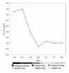
After 60 days of steroid treatment, oral prednisolone acetate was tapered off to 50 mg (30 mg/20 mg) daily. After ninety days of steroid treatment, oral prednisolone was tapered off to 15 mg daily, and one month later oral prednisolone was tapered off to 10 mg daily. The 10 mg dose of oral prednisolone was maintained for 4 months, after which time the current dose of 5 mg daily began (Fig. 1).
During a 10-month follow-up examination, the patient's thyroid function test was determined to be within normal limits with PTU therapy, and no neurological symptoms were present with tapered corticosteroid therapy. Presently, the patient is a follow-up outpatient without any special symptoms and has been taking oral prednisolone acetate 5 mg #1, PTU 100 mg #2, propranolol HCl 40 mg #2, and clonazepam 1.0 mg #2.
In summary, the patient showed prominent choreic movement in the left arm and distal left leg prior to steroid treatment. Continuous choreic movements in her left foot, left hand, and perioral area disabled her daily life. After 7 days of steroid treatment, a follow up picture was taken. The choreic movements presenting in her extremities and perioral area had resolved and she has not had any involuntary movements since that time.
Discussion
Chorea is a state of involuntary movement, which starts suddenly and is characterized by rapid movements; however, it is also forms a complex of cooperative movement[11~13]. Lesions of the central nervous system and peripheral nervous system can cause chorea[14]. When associated with thyroid disease, chorea is most commonly observed in Hashimoto's thyroiditis[15].
There have been four reported cases of chorea that have presented with Graves' disease[16~18]. In one of these cases, choreic movements and thyroid hormones were not controlled by steroid treatment combined with anti-thyroid medication. Radioiodine therapy and total thyroidectomy, respectively, were necessary. One patient showed a relapsing episode of chorea. In two of the patients, the recurrence of chorea was observed with tapering of corticosteroid therapy. However, the mechanisms underlying this phenomenon have not been clearly defined.
The diagnosis of Graves' disease in our patient was confirmed by laboratory tests and RAIU tests. Since other causes of chorea were excluded and choreic movements improved concurrently with the decrease of anti-microsomal antibody titer, we could make a diagnosis of chorea secondary to Graves' disease in our patient based on confirmed parameters. Treatment of chorea associated with autoimmune thyroid disease has mainly consisted of immunomodulatory agents (mostly steroids) and/or thyroid-modulating agents, as well as antichoreic drugs. There are no definite guidelines for steroid treatment in chorea secondary to Graves' disease. But it is considered that steroid treatment should be tapered off carefully according to the patient's symptoms.
Until the present study, the pathogenesis of chorea secondary to Graves' disease had not been established. Some studies suggest increased sensitivity of dopamine receptors and increased levels of catecholamines in the brain and neuromuscular system. Other studies have implied that a coincident abnormality of the basal ganglia must also be present in hyperthyroid patients who develop chorea. In the only other reported patient with hyperthyroidism and ataxia, the ataxia developed well in advance of the hyperthyroidism. This would suggest that the ataxia was related to underlying autoimmune disease rather than the direct effect of the hyperthyroidism.
In a previous case report, an eruption of chorea was observed in a patient without autoimmune thyroid disease, but with hypothyroidism, despite the administration of thyroid hormone[19]. Additionally, in chorea secondary to Graves' disease, the recurrence of chorea occurred when hyperthyroidism recurred.
On the other hand, autoimmune system dysfunction in encephalopathy associated with autoimmune thyroid disease (EAATD) could be considered at the onset of chorea, as demonstrated in many other autoimmune diseases[20]. There have been several case reports about EAATD. Most of these reports involved Hashimoto's thyroiditis, and chorea occurred mainly during the hypothyroid state. Upon suspicion of EAATD, corticoid administration should be started immediately in order to see dramatic improvement.
Because chorea secondary to Graves' disease occurred even in normal thyroid function status in previous reports and choreic movements improved following the initiation of steroid treatment before the recovery of hyperthyroidism in this patient, we suggest that the occurrence of chorea secondary to Graves' disease could be dominantly associated with autoimmune dysfunction rather than with increased levels of thyroid hormone. Future research examining changes in basal ganglia sensitivity to thyroid hormone or disorder in autoimmune reaction may be warranted in the future.
 XML Download