Abstract
Tumor-induced osteomalacia (TIO), a paraneoplastic disease, is characterized by hypophosphatemia, and caused by renal phosphate wasting inappropriately, normal or decreased 1, 25(OH)2D3 production, and defective calcification of cartilage and bone. Because the removal of the responsible tumor normalizes phosphate metabolism, unidentified humoral phosphaturic factors (phosphatonin) are believed to be responsible for this syndrome. These factors include fibroblast growth factor (FGF)-23, secreted frizzled-related protein-4 and matrix extracellular phosphoglycoprotein. However, no case of TIO producing FGF-23 has been clearly reported in Korea. Herein, a case of TIO producing FGF-23 in a 45-year-old woman is reported. The patient presented with a large tumor on her buttock, with severe bone and muscle pain. A histological examination of the tumor revealed a mixed connective tissue tumor, consisting of deposition of calcified materials and surrounding primitive spindle cells, with prominent vascularity. Whether FGF-23 is a secreted factor, as well as its levels of expression in tumors were investigated. An immunohistochemical study showed the tumor cells to be FGF-23 positive. Furthermore, the levels of serum FGF-23 were extremely high and an RT-PCR analysis, using total RNA from the tumor, revealed the abundant expression of FGF-23 mRNA. After removal of the tumor, all the biochemical and hormonal abnormalities disappeared, with marked symptomatic improvement.
Figures and Tables
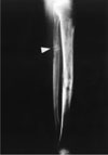
Fig. 1
Plain x-ray film of both lower legs at initial admission (1990). Radiography showing radiolucent line and loss of cortical continuity (pseudofracture) of the upper diaphysis of the left fibula.

Fig. 2
Pelvic CT in patient who was presented with a large buttock mass shows large dumbbell shape soft tissue mass posterior to rectum and left side buttock.

Fig. 3
H & E and immunohistochemical staining for FGF-23 in tumor. A, Tumor cells are composed of small bland spindle to ovoid cells that produce a distinctive eosinophillic smudge matrix with deposition of calcified materials (grungy pattern). H&E, × 400; B, The prominent branching vascular patterns mimic those seen in hemangiopericytoma/solitary fibrous tumor. H&E, × 400; C, Positive staining of FR tumor with antiFGF-23 antibody under high power. Staining was cytoplasmic and granular. Arrows indicate the presence of immunostained cells (brown staining), × 400.

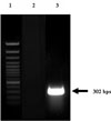
Fig. 4
RT-PCR analysis of FGF-23 mRNA in tumor. Agarose gels with separated PCR products are shown. Lanes: 1, molecular weight markers; 2, adrenal adenoma tissue from a patient with primary aldosteronism; 3, tumor tissue. The predicted sizes of the PCR products in base pairs are indicated to the right of the panel.

References
1. Shimada T, Mizutani S, Muto T, Yoneya T, Hino R, Takeda S, Takeuchi Y, Fujita T, Fukumoto S, Yamashita T. Cloning and characterization of FGF23 as a causative factor of tumor-induced osteomalacia. Proc Natl Acad Sci U S A. 2001. 98:6500–6505.
2. Kronenberg HM. NPT2a--the key to phosphate homeostasis. N Engl J Med. 2002. 347:1022–1024.
3. Schiavi SC, Kumar R. The phosphatonin pathway: new insights in phosphate homeostasis. Kidney Int. 2004. 65:1–14.
4. McCrane R. Osteomalacia with Looser's node (Milk-man's syndrome) due to raised resistance to vitamin D aquired about the age of 15 years. Q J Med. 1947. 16:33–46.
5. Prader A, Illig R, Uehlinger E, Stalder G. [Rickets following bone tumor.]. Helv Paediatr Acta. 1959. 14:554–565.
6. Folpe AL, Fanburg-Smith JC, Billings SD, Bisceglia M, Bertoni F, Cho JY, Econs MJ, Inwards CY, Jan de, Mentzel T, Montgomery E, Michal M, Miettinen M, Mills SE, Reith JD, O'Connell JX, Rosenberg AE, Rubin BP, Sweet DE, Vinh TN, Wold LE, Wehrli BM, White KE, Zaino RJ, Weiss SW. Most osteomalacia-associated mesenchymal tumors are a single histopathologic entity: an analysis of 32 cases and a comprehensive review of the literature. Am J Surg Pathol. 2004. 28:1–30.
7. Jan de Beur SM, Streeten EA, Civelek AC, McCarthy EF, Uribe L, Marx SJ, Onobrakpeya O, Raisz LG, Watts NB, Sharon M, Levine MA. Localisation of mesenchymal tumours by somatostatin receptor imaging. Lancet. 2002. 359:761–763.
8. Narvaez J, Rodriguez-Moreno J, Moragues C, Campoy E, Clavaguera T. Br J Rheumatol. 1996. 35:598–600.
9. Henneman PH, Dempsey EF, Carroll EL, Henneman DH. Metabolism. 1962. 11:103–116.
10. Daniel S, Ofer BI, Alicia N, Amira B, Rachel BS, Ibrahim S, Lael AB. Semin Arthritis Rheum. 1995. 25:35–46.
11. Harvey JM, Gray C, Belchetz PE. Clin Endocrinol. 1992. 37:379–384.
12. The Consortium ADHR. Autosomal dominant hypophosphataemic rickets is associated with mutations in FGF23. Nat Genet. 2000. 26:345–348.
13. The Consortium HYP. A gene (PEX) with homologies to endopeptidases is mutated in patients with X-linked hypophosphatemic rickets. The HYP Consortium. Nat Genet. 1995. 11:130–136.
14. Campos M, Couture C, Hirata IY, Juliano MA, Loisel TP, Crine P, Juliano L, Boileau G, Carmona AK. Human recombinant endopeptidase PHEX has a strict S1' specificity for acidic residues and cleaves peptides derived from fibroblast growth factor-23 and matrix extracellular phosphoglycoprotein. Biochem J. 2003. 373:271–279.
15. Guo R, Liu S, Spurney RF, Quarles LD. Analysis of recombinant Phex: an endopeptidase in search of a substrate. Am J Physiol Endocrinol Metab. 2001. 281:E837–E847.
16. Weber TJ, Liu S, Indridason OS, Quarles LD. Serum FGF23 levels in normal and disordered phosphorus homeostasis. J Bone Miner Res. 2003. 18:1227–1234.
17. Gupta A, Winer K, Econs MJ, Marx SJ, Collins MT. FGF-23 is elevated by chronic hyperphosphatemia. J Clin Endocrinol Metab. 2004. 09. 89(9):4489–4492.
18. Sato T, Tominaga Y, Ueki T, Goto N, Matsuoka S, Katayama A, Haba T, Uchida K, Nakanishi S, Kazama JJ, Gejyo F, Yamashita T, Fukagawa M. Total parathyroidectomy reduces elevated circulating fibroblast growth factor 23 in advanced secondary hyperparathyroidism. Am J Kidney Dis. 2004. 09. 44(3):481–487.
19. Kobayashi K, Imanishi Y, Miyauchi A, Onoda N, Kawata T, Tahara H, Goto H, Miki T, Ishimura E, Sugimoto T, Ishikawa T, Inaba M, Nishizawa Y. Regulation of plasma fibroblast growth factor 23 by calcium in primary hyperparathyroidism. Eur J Endocrinol. 2006. 01. 154(1):93–99.
20. Imel EA, Peacock M, Pitukcheewanont P, Heller HJ, Ward LM, Shulman D, Kassem M, Rackoff P, Zimering M, Dalkin A, Drobny E, Colussi G, Shaker JL, Hoogendoorn EH, Hui SL, Econs MJ. Sensitivity of fibroblast growth factor 23 measurements in tumor-induced osteomalacia. J Clin Endocrinol Metab. 2006. 91:2055–2061.
21. Yamazaki Y, Okazaki R, Shibata M, Hasegawa Y, Satoh K, Tajima T, Takeuchi Y, Fujita T, Nakahara K, Yamashita T, Fukumoto S. Increased circulatory level of biologically active full-length FGF-23 in patients with hypophosphatemic rickets/osteomalacia. J Clin Endocrinol Metab. 2002. 87:4957–4960.
22. Ryan EA, Reiss E. Oncogenous osteomalacia. Review of the world literature of 42 cases and report of two new cases. Am J Med. 1984. 77:501–512.


 PDF
PDF ePub
ePub Citation
 Print
Print


 XML Download