

 PDF
PDF ePub
ePub Citation
 Print
Print


Abstract
Purpose
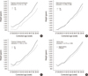
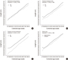
Study purpose were to describe growth patterns of premature infants in weight, length and head circumference from birth to 40th week of corrected ages (CA) and to explore factors affecting patterns.
Methods
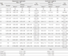
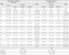
A longitudinal descriptive study was conducted with 267 premature infants. They were categorized into 2 groups; GA group with measurements at birth and the CA group with measurements at CA, which was categorized into 3 groups (group 1-3) by WHO guideline for gestational age (GA) at birth.
Results
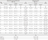
GA group presented greater measures in all than CA group at same week of life. Among CA groups, group 3 showed the highest measurements, up to 37 weeks of life, though this disappeared at 38-40 weeks. Reversely, group 1 revealed the highest growth rates in all measures, followed by group 2 and group 3. Significant interaction was observed in all measures between week of life and any type of groups.
Conclusion
Higher measures in GA group, as well group 3 among CA groups, supported the superiority of intra-uterine environment overriding quality of regimen from NICU. Regardless of growth acceleration, smaller infants remain smaller, indicating that intra-uterine thrifty phenotype may continue at least up to the 40th week of CA.
Figures and Tables

References
1. Adair LS. Size at birth and growth trajectories to young adulthood. American Journal of Human Biology. 2007. 19:327–337. doi: 10.1002/ajhb.20587.
2. Ahn Y. Assessment of gestational age using an extended New Ballard examination in Korean newborns. Journal of Tropical Pediatrics. 2008. 54:278–281. doi: 10.1093/tropej/fmm120.
3. Ahn Y, Garruto RM. Weight variation by sex and nature of risk factors in high-risk infants: An evolutionary perspective. Collegium Antropologicum. 2007. 31:937–941.
4. Babson SG, Benda GI. Growth graphs for the clinical assessment of infants of varying gestational age. Journal of Pediatrics. 1976. 89:814–820. doi: 10.1016/S0022-3476(76)80815-3.
5. Campbell MK, Mottola MF. Recreational exercise and occupational activity during pregnancy and birth weight: A case-control study. American Journal of Obstetrics and Gynecology. 2001. 184:403–408. doi: 10.1067/mob.2001.109392.
6. Dawodu A, Bener A, Koutouby GA, Varady E, Abdulrazzaq Y. Size at birth in a rapidly developing economy: Intrauterine growth pattern of UAE infants. Annals of Human Biology. 2008. 35:615–623. doi: 10.1080/03014460802385439.
7. de Onis M, Onyango AW, Borghi E, Garza C, Yang H. WHO Multicentre Growth Reference Study Group. Comparison of the world health organization (WHO) child growth standards and the National Center for Health Statistics/WHO international growth reference: Implications for child health programmes. Public Health Nutrition. 2006. 9:942–947. doi: 10.1017/PHN20062005.
8. Euser AM, de Wit CC, Finken MJ, Rijken M, Wit JM. Growth of preterm born children. Hormone Research. 2008. 70:319–328. doi: 10.1159/000161862.
9. Gardner SL, Carter BS, Enzman-Hines M, Hernandes JA, editors. Merenstein & Gardner's handbook of neonatal intensive care. 2011. New York: Mosby.
10. Goldenberg RL, Culhane JF, Iams JD, Romero R. Epidemiology and causes of preterm birth. Lancet. 2008. 371(9606):75–84. doi: 10.1016/S0140-6736(08)60074-4.
11. Hales CN, Ozanne SE. The dangerous road of catch-up growth. Journal of Physiology. 2003. 547:5–10. doi: 10.1113/jphysiol.2002.024406.
12. 2007 Korean national growth charts: Review of developmental process and outlook. Korea Centers for Disease Control and Prevention. 2008. 04. 23. Retrieved August 31, 2010. from http://www.cdc.go.kr/kcdchome/jsp/home/information/had/INFOHAD0001Detail.jsp?menuid=100053&appid=kcdchome&content=/contents/information/had/b/4182_view.html.
13. Statistics of birth in whole nation & cities and provinces (1998-2009). Korean Statistical Information Service. 2009. Retrieved March 12, 2011. from http://kosis.kr/abroad/abroad_01List.jsp.
14. Magann EF, Evans SF, Weitz B, Newnham J. Antepartum, intrapartum, and neonatal significance of exercise on healthy low-risk pregnant working women. Obstetrics and Gynecology. 2002. 99:466–472.
15. Olsen IE, Groveman SA, Lawson ML, Clark RH, Zemel BS. New intrauterine growth curves based on United States data. Pediatrics. 2010. 125:e214–e224. doi: 10.1542/peds.2009-0913.
16. Ong KK. Catch-up growth in small for gestational age babies: Good or bad? Current Opinion in Endocrinology, Diabetes, and Obesity. 2007. 14:30–34. doi: 10.1097/MED.0b013e328013da6c.
17. Prada JA, Tsang RC. Biological mechanisms of environmentally induced causes of IUGR. European Journal of Clinical Nutrition. 1998. 52:S21–S27. discussion S27-S28.
18. Rao SC, Tompkins J. Growth curves for preterm infants. Early Human Development. 2007. 83:643–651. doi: 10.1016/j.earlhumdev.2007.07.008.
19. Remacle C, Bieswal F, Reusens B. Programming of obesity and cardiovascular disease. International Journal of Obesity. 2004. 28:S46–S53. doi: 10.1038/sj.ijo.0802800.
20. Ross SA, Milner JA. Epigenetic modulation and cancer: Effect of metabolic syndrome? American Journal of Clinical Nutrition. 2007. 86:s872–s877.
21. Srinivas SK, Edlow AG, Neff PM, Sammel MD, Andrela CM, Elovitz MA. Rethinking IUGR in preeclampsia: Dependent or independent of maternal hypertension? Journal of Perinatology. 2009. 29:680–684. doi: 10.1038/jp.2009.83.
22. Stinson S. Stinson S, Bogin B, Huss-Ashmore R, O'Rourke D, editors. Growth variation: Biological and cultural factors. Human biology: An evolutionary and biocultural perspectives. 2000. New York, NY: Wiley-LISS;425–464.
23. Thureen PJ. The neonatologist's dilemma: Catch-up growth or beneficial undernutrition in very low birth weight infants-what are optimal growth rates? Journal of Pediatric Gastroenterology and Nutrition. 2007. 45:S152–S154. doi: 10.1097/01.mpg.0000302962.08794.62.
24. Wells JC. The programming effects of early growth. Early Human Development. 2007. 83:743–748. doi: 10.1016/j.earlhumdev.2007.09.002.
25. WHO Multicentre Growth Reference Study Group. WHO child growth standards based on length/height, weight and age. Acta paediatrica. Supplementum. 2006. 450:76–85. doi: 10.1080/08035320500495548.
 XML Download