

 PDF
PDF ePub
ePub Citation
 Print
Print


INTRODUCTION
The public health burden of diabetic nephropathy has increased rapidly along with the world-wide increase in the prevalence of type 2 diabetes. Diabetic nephropathy deteroriates quality of life and also decreases life expectancy. Microalbuminuria is a predictor of overt proteinuria in patients with diabetes [1], as well as being a risk factor for cardiovascular disease in patients with type 2 diabetes and in normal subjects [2]. Although poor glycemic control and high blood pressure are known to increase the risk of microalbuminuria in patients with type 2 diabetes [3-5], little is known regarding other factors that predispose these individuals to the development of microalbuminuria.
Hyperhomocysteinemia occurs frequently in patients with renal failure [6]. As proper renal function is crucial to homocysteine metabolism [7], it is generally believed that hyperhomocysteinemia in renal failure is secondary to decreased renal function. On the other hand, recent studies in animals showed that hyperhomocysteinemia induces glumerulosclerosis and podocyte injury [8-12]. It has also been reported that hyperhomocysteinemia precedes the development of overt proteinuria in patients with type 2 diabetes [13]. These findings suggest that hyperhomocysteinemia may play a pathogenic role in the genesis of diabetic nephropathy. However, it is not yet clear whether hyperhomocysteinemia can be a risk factor for early diabetic nephropathy. We therefore assessed whether hyperhomocysteinemia is an independent risk factor for the development of microalbuminuria in patients with type 2 diabetes.
METHODS
Study protocol
This study was performed in patients with type 2 diabetes attending a diabetes clinic of a university hospital (the Asan Medical Center) in Seoul, South Korea. From January 2002 to December 2004, we recruited 1,226 patients with type 2 diabetes and normoalbuminuria at the time of registry and with urine collection. Patients were excluded if any one of the following conditions was present: plasma creatinine > 1.2 mg/dL, congestive heart failure, or a major concurrent illness such as cancer or end-stage pulmonary or liver disease. After obtaining written informed consent from each individual and the approval of our institutional review committee, we obtained baseline blood samples from each patient, as well as recording patients age, sex, blood pressure, duration of diabetes and other clinical information. Blood samples were centrifuged and stored at -70℃.
At baseline, patients collected timed overnight urine samples 2 or 3 times [5], and their baseline albumin excretion rate was determined by radioimmunoassay (Beckman Coulter, Galway, Ireland). Individuals were considered normoalbuminuric if urinary albumin excretion (UAE) was consistently under 20 µg/min. Patients were followed every 3 months. At each visit, blood pressure, plasma glucose, lipid profiles, and HbA1C were measured. UAE was measured twice yearly from timed overnight urine collections. If the UAE value exceeded 20 µg/min, it was measured twice more at 3-month intervals. Persistent microalbuminuria was defined as UAE > 20 µg/min on at least two of the three measurements.
By June 2007, we collected data on 887 patients who had been followed for more than 18 months (mean follow up, 36.0 ± 11.7 months; range, 18 to 76 months). Among these, 76 subjects developed microalbuminuria during the follow-up period, and we defined them as cases. For each case, we randomly and individually selected two controls who were matched on age and sex.
Laboratory analyses
Fasting plasma glucose, HbA1C, C-peptide, creatinine, high sensitivity C-reactive protein (hsCRP), total cholesterol, triglycerides, high density lipoptoein cholesterol (HDL-C), low density lipoptoein cholesterol (LDL-C) and free fatty acid concentrations were measured by standard procedures. Homocysteine concentrations in the stored plasma samples were measured by competitive immunoassay analyzed on the ADVIA Centaur (Bayer Diagnostics, Tarrytown, NY, USA). Estimated glomerular filtration rate (GFR) was calculated using Cockroft & Gault equation. Matched case-control pairs were handled identically and assayed in random order in the same analytical run.
Statistical analyses
Results are reported as mean ± standard deviation or median with interquartile range. Statistical analysis was performed using SPSS software (version 12.0; SPSS Inc., Chicago, IL, USA). Student's t-test or Mann Whitney U test was used for continuous variables, and the χ2 test for analysis of discrete variables, with all P values reported for two-sided tests. Values of P < 0.05 were considered significant.
To determine the associations of potential determinants with the development of microalbuminuria, we performed multiple logistic regression analysis with backward elimination of variables, using seven variables that may be of biological significance (i.e., baseline homocysteine and estimated GFR levels, baseline UAE, follow-up duration, mean systolic blood pressure and HbA1C levels during follow-up, past and present cardiovascular disease). The presence of microalbuminuria at follow-up was the dependent variable and potential determinants for development of microalbuminuria were independent variables. Variables having skewed distribution such as baseline plasma homocysteine concentrations, and follow-up duration were natural log (Ln)-transformed before analysis. Values of P < 0.05 were considered significant. The dose-response relationship for homocysteine was determined by calculating odds ratios for developing microalbuminuria for several total homocysteine concentration ranges (9.1 to 14.0, 14.1 to 19.0, and > 19.0 µmol/L) with total homocysteine concentrations < 9.1 µmol/L as reference [14].
RESULTS
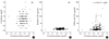
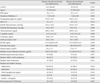
Table 1 shows the baseline clinical characteristics of patients who did and did not develop microalbuminuria. We observed no significant between-group differences in baseline age, sex, body mass index (BMI), duration of diabetes, fasting plasma glucose, systolic and diastolic blood pressure, UAE, and HbA1C, total cholesterol, C-peptide, creatinine, estimated GFR, hsCRP, free fatty acid concentrations, prevalence of hypertension, the use of antihypertensive or anti-diabetic medications and past and present cardiovascular disease history. There was no statistically significant correlation between Ln plasma homocysteine level and baseline creatinine levels or UAE (Fig. 1A and B).
Baseline concentrations of plasma homocysteine, however, were significantly higher among patients who developed microalbuminuria than among patients who were normoalbuminuric during the follow-up period (11.8 ± 3.7 µmol/L vs. 10.3 ± 3.1 µmol/L, P = 0.003). There was a significant positive correlation between baseline Ln plasma homocysteine levels and follow-up UAE (Fig. 1C).
There were no significant between group differences in duration of follow up, mean systolic blood pressure, mean diastolic blood pressure, and mean total cholesterol (Table 2). However, mean HbA1C during follow-up were significantly higher among patients who developed microalbuminuria than among patients who remained normoalbuminuric (7.5 ± 0.9% vs. 8.0 ± 1.2%, P = 0.001).
Multivariate binary logistic regression analysis with backward elimination of variables was performed using baseline Ln plasma homocysteine and estimated GFR levels, baseline UAE, Ln follow-up duration, mean systolic blood pressure and mean HbA1C levels during follow-up, and past and present cardiovascular disease history as independent variables, and microalbuminuria development as the dependent variable. Mean HbA1C (odds ratio [OR], 1.750; range, 1.297 to 2.362; P < 0.001) and Ln homocysteine levels (OR, 5.167; range, 1.898 to 14.067; P = 0.001) were independent predictors of microalbuminuria development (Table 3).
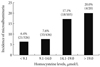
We divided the total subjects in whom baseline total homocysteine concentrations were measured (n = 887) into those with baseline homocysteine concentrations of < 9.1, 9.1 to 14.0, 14.1 to 19.0, and > 19.0 µmol/L. The cumulative incidence of microalbuminuria in these 4 groups of patients was 6.4% (21/326), 7.6% (33/436), 17.1% (18/105), and 20.0% (4/20), respectively (P value for trend = 0.001, Fig. 2). After adjustment for age, sex, baseline HbA1C and plasma creatinine concentrations, the odds ratios for microalbuminuria, relative to baseline homocysteine concentrations of < 9.1 µmol/L, were 1.3 (95% confidence interval [CI], 0.7 to 2.4), 3.6 (95% CI, 1.8 to 7.3), and 4.6 (95% CI, 1.4 to 15.4) for baseline homocysteine concentrations of 9.1 to 14.0, 14.1 to 19.0, and > 19.0 µmol/L, respectively. When homocysteine level categories were replaced by homocysteine concentration as a continuous variable, a 5 µmol/L increase in homocysteine concentration was associated with a 1.8-fold (95% CI, 1.3 to 2.5; P < 0.001) increase in the risk of developing microalbuminuria, after adjustment for age, sex, baseline HbA1c and creatinine levels.
DISCUSSION
In this study, we show that hyperhomocysteinemia is an independent risk factor for the development of microalbuminuria in patients with type 2 diabetes. Baseline plasma homocysteine concentrations were significantly higher in normoalbuminuric patients who developed microalbuminuria during follow-up than in normoalbuminuric patients who did not develop microalbuminuria. These findings are in agreement with previous cross-sectional and longitudinal studies [13,15], which showed associations between plasma homocysteine levels and overt proteinuria in diabetic patients. Although homocysteine concentrations have been found to be associated with microalbuminuria development in non-diabetic subjects [14], to our knowledge, the present study is the first to show an association between plasma homocysteine concentration and development of microalbuminuria in diabetic individuals. Although the previous study by Jager et al. was unable to find a relationship between hyperhomocysteinemia and microalbuminuria developement in diabetic patients [14], our results showed the significant relationship in patients with type 2 diabetes because of a larger sample size.
Kidney function is critical for homocysteine clearance [7], and hyperhomocysteinemia occurs frequently in patients with renal failure [6,16]. Thus it can be asked whether the association between plasma homocysteine levels and urinary albumin excretion is attributed to associated changes in renal function [16,17]. In our study, however, only patients who were normoalbuminuric at baseline were included. Although we did not measure glomerular filtration rate in all of these patients, renal function is known to deteriorate following (micro)albuminuria [18,19]. All of our subjects had normal plasma creatinine levels (below 1.2 mg/dL), and there was no significant correlation between baseline Ln plasma homocysteine level and baseline UAE or baseline plasma creatinine concentration. On the other hand, follow-up UAE level was significantly correlated with baseline Ln plasma homocysteine concentration.
Homocysteine has been shown to cause vascular disease and endothelial damage [20-22]. In addition, recent studies suggested that homocysteine is toxic to kidney tissues. Dietinduced chronic hyperhomocysteinemia could induce arterial and arteriolar thickening, and tubulointerstitial and podocyte injury in the kidney [8,9]. Regarding the mechanism of homocysteine-induced glomerular injury, homocysteine was shown to activate MAP kinases and to induce endoplasmic reticulum stress in cultured mesangial cells [10]. It was also shown that homocysteine stimulates ceramide-mediated redox signaling [11], and increases monocyte chemoattractant protein-1 expression in the kidney via nuclear factor-kappaB activation [12]. These studies suggest that hyperhomocysteinemia may play a causative role in early renal injury, in addition to being a marker of impaired renal function [16,17].
Poor glycemic control and high blood pressure are known to increase the risk of microalbuminuria in patients with type 2 diabetes [3-5]. As expected, we found significant differences between cases and controls in mean HbA1C levels during follow-up. In addition, multivariate logistic regression analyses showed that basal Ln plasma homocysteine levels remained a significant independent variable, even after correcting for mean HbA1C levels and other important variables. We also found that plasma homocysteine concentration > 14.0 µmol/L was associated with a marked increase in the risk of microalbuminuria. For each 5 µmol/L increase in homocysteine concentration, the risk of developing microalbuminuria increased by about 80%. This is in line with previous studies showing 1.3 to 1.6 fold increase per 5 µmol/L increase in homocysteine concentration in the risk for microalbuminuria in non-diabetic subjects [23] and of overt proteinuria in diabetic subjects [13].
Although microalbuminuria in type 1 diabetes is regarded as an early manifestation of diabetic nephropathy, the meaning of microalbuminuria in type 2 diabetes is more complex. The metabolic syndrome and obesity, which frequently accompany type 2 diabetes, predispose these individuals to microalbuminuria [24], and plasma homocysteine level is frequently increased in patients with metabolic syndrome [25,26].
In the present study, 125 among 887 patients had marked hyperhomocysteinemia (> 14 µmol/L) and increased risk for developing microalbuminuria. Since homocysteine has been shown to cause cardiovascular disease and endothelial damage [20-22], hyperhomocysteinemia in diabetic patients may lead to endothelial dysfunction in systemic and renal blood vessels, and predispose these patients to increased risks of both cardiovascular disease and microalbuminuria.
In our study, we used nested cohort design with a relatively small number of patients attending a single center. For many research questions, the nested case-control design potentially gives significant reductions in time, cost, and effort of data collection and analysis, compared with the full cohort approach, with a relatively minor loss in statistical efficiency [27,28]. However, future studies with a larger population and longer duration of follow-up are needed to provide more definitive conclusion.
In summary, we have shown that hyperhomocysteinemia is associated with a higher incidence of microalbuminuria in patients with type 2 diabetes mellitus. Further studies in larger populations are needed to confirm these findings, and to test whether lowering of plasma homocysteine by dietary manipulation [29] is helpful for patients at risk for diabetic nephropathy.


 XML Download