

 PDF
PDF ePub
ePub Citation
 Print
Print


Introduction
The incidence of cardiovascular disease (CVD) in women increases with age1-6). CVD occurrence is distinct in both men and women, and onset begins about ten years later in women than men. Myocardial infarction is uncommon until women reach their sixth decade; and although women below the age of fifty rarely develop CVD, by age seventy their incidences reach equally to that of men7). This suggests that estrogen deficiency contributes to the rise in CVD risk. The decrease in estrogen is also related to elevated low density lipoprotein (LDL) cholesterol levels1,3-6).
Postmenopausal status is associated with a 60% increased risk of metabolic syndrome8). Characterized by the association of different metabolic syndrome risk factors such as glucose intolerance, abdominal obesity, dyslipidemia, and hypertension, metabolic syndrome is an important determinant of cardiovascular morbidity and mortality9). The etiology is unknown, but is hypothesized to be caused by several factors. Many believe that underlying pathophysiology of metabolic syndrome is related to increased visceral obesity and insulin resistance10-12). There are considerable age-dependent rise of both insulin resistance and visceral obesity, especially in women these factors are further altered by menopause. In essence, each factor increases progressively after menopause.
Patients and Methods
1. Study Population
We conducted a cross-sectional study. A total of 1,926 women, who had visited the health examination center in the Wonju Christian Hospital at Yonsei University Wonju College of Medicine between March 2005 and February 2006, were included in this study. They didn't take any hormonal medication like estrogen, progesterone, or mixtures in the past medical history. They were divided into three groups according to their menstrual stage. We defined the perimenopause as 3 to 11 months of amenorrhea or changes in menstrual regularity (either shortening or lengthening of time between menses) among women aged 45 to 55 years14) and the menopause is defined after 12 months of amenorrhea15). There were 1,274 premenopausal women with a normal menstrual history, 205 perimenopausal women and 447 postmenopausal women. We excluded women with pregnancy, 1 year or less post-parturition, use of hormone replacement therapy, acute illness, oral contraceptive use, prior hysterectomy or oophorectomy.
2. Data Collection and Assays
The height and weight of each patient was examined with light clothing in the morning. Body mass index (BMI) was calculated as kg/m2 and blood pressure was measured with a mercury sphygmomanometer after the patient had been seated at rest for 10 minutes. Waist circumference was measured midway between the lateral lower rib margin and the anterior superior iliac crest according to World Health Organization (WHO) criteria.
All venous blood samples were drawn in the morning after 10 hours of overnight fasting. Total cholesterol, triglyceride, HDL cholesterol, glucose, ALT, aspartate aminotransferase (AST), GGT, and uric acid were measured using an autoanalyser (Hitachi, Tokyo, Japan). White blood cell counts were measured using an autoanalyser (Bayer, New York). High sensitive C-reactive protein (hsCRP) levels were determined using the latex aggregation method (Roche, Indianapolis, IN).
3. Definition of Metabolic Syndrome
We adopted the NCEP Expert Panel on the Detection, Evaluation, and Treatment of High Blood Cholesterol in Adults (Adult Treatment Panel III; ATP III) as the criteria for metabolic syndrome in our study. But we adopted the Korean Society for the Study of Obesity (KSSO) criteria about waist circumference for women16). Criteria were as follows: central waist circumference ≥ 85 cm, triglyceride ≥ 1.7 mmol/L (150 mg/dL), HDL cholesterol < 1.29 mmol/L (50 mg/dL), blood pressure ≥ 130 mmHg (systolic) or ≥ 85 mmHg (diastolic), and fasting plasma glucose ≥ 5.6 mmol/L (100 mg/dL). The subjects were classified as having metabolic syndrome if they possessed three or more of the five components. The number of metabolic syndrome components determined the metabolic syndrome score.
4. Statistical Analysis
Results were expressed as the mean ± standard deviation, and differences were considered significant at a P value < 0.05. Multivariate ANOVA was used to compare the three groups, and correlations between variables were calculated using the Spearman test. Logistic regression analyses were used to obtain an odds ratio for waist circumference, triglyceride, HDL cholesterol, blood pressure, and fasting plasma glucose. All analyses were performed by the program SPSS for Windows (version 12.0, SPSS Inc., Chicago, IL).
Results
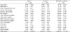
Table 1 shows the baseline clinical and biochemical characteristics of the women in the study. The average age was 43.8 years, and the patients classified with metabolic syndrome numbered 219 (11.3% overall prevalence).
1. Comparison of Anthropometric and Biochemical Characteristics Among the Groups
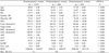
Of the 1,926 women, 66.2% were classified as premenopausal, 10.6% as perimenopausal, and 23.2% as postmenopausal. The prevalence of metabolic syndrome was 7.1% in premenopause, 9.8% in perimenopause, and 24.2% in postmenopause. Postmenopausal women were characterized by a higher body mass index, waist circumference, and blood pressure as compared to premenopausal women (P < 0.001). Postmenopausal women also presented with higher fasting glucose, total cholesterol, triglyceride, uric acid, LDL cholesterol, white blood cell (WBC) count, and lower HDL cholesterol than premenopausal women (Table 2).
2. Relationships Between Metabolic Syndrome Score and Various Clinical and Biochemical Parameters
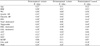
A strong positive correlation was noted between metabolic syndrome score and body mass index, waist circumference, systolic blood pressure, and triglyceride in premenopausal women (0.56, 0.56, 0.51, and 0.53). In perimenopausal women waist circumference, systolic blood pressure, and triglyceride were the strong positive correlation with the metabolic syndrome score (0.51, 0.60, and 0.54). In postmenopausal women, body mass index, waist circumference, and systolic blood pressure were the strong positive correlation with metabolic syndrome score (0.51, 0.56, and 0.51). A negative correlation was noted between the metabolic syndrome score and HDL cholesterol in premenopausal, perimenopausal, and postmenopausal women. Age was the positive correlation in premenopausal and postmenopausal women according to increase of metabolic syndrome score. Other parameters of metabolic syndrome, including ALT, GGT, uric acid, and WBC were also positively correlated with metabolic syndrome score (Table 3).
3. Relative Risk Between Metabolic Syndrome and Its Components in Menopausal Transition
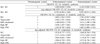
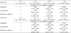
Based on a multiple regression analysis, a risk of metabolic syndrome was increased in postmenopausal women. Triglyceride, blood pressure, and waist circumference showed increased relative risk in perimenopausal women compared with premenopausal women (Table 4). However, when these components were adjusted by age, only triglyceride was significantly associated with menopausal transition (OR 1.517 [95% CI 1.014-2.269]; P = 0.042 in premenopausal women, OR 1.573 [95% CI 1.025-2.414] P = 0.038 in postmenopausal women).
4. Relative Risk for Metabolic Syndrome According to Serum-glutamyl Transpeptidase and Alanine Aminotransferase Concentrations
Higher concentrations of GGT and ALT were risk factors for metabolic syndrome, although their levels were within normal range, and these relationships are independent of age (Table 5).
Discussion
In our study, the prevalence of metabolic syndrome increased throughout the menopausal transition. The relative risk of metabolic syndrome components increased in postmenopause. The prevalence of metabolic syndrome was 11.3% in all women, 7.1% in premenopausal women, 9.8% in perimenopausal women, and 24.2% in postmenopausal women. These results are similar to other studies8,12,17) even though we utilized NCEP ATP III criteria (waist circumference ≥ 85 cm) to define metabolic syndrome.
Metabolic syndrome is a formidable risk factor for the development of type 2 diabetes, and the risk for diabetes is nearly fivefold in patients with metabolic syndrome18). Diabetes also increases the risk of coronary artery disease and stroke by two to four fold19,20). This study demonstrated an increase in fasting plasma glucose throughout the menopausal transition, and showed positive correlation between the metabolic syndrome score and elevated fasting glucose level in each group.
Metabolic syndrome is also a strong risk factor for CVD, and incidences in women increase with age1-6), followed by elevated total cholesterol, LDL cholesterol, and apolipoprotein B1,6). In this study total cholesterol, LDL cholesterol, and triglyceride increased in postmenopausal women and HDL cholesterol level decreased in postmenopausal women. Triglyceride and HDL cholesterol were associated with metabolic syndrome score in each group. However the total cholesterol and LDL cholesterol correlated with metabolic syndrome score in premenopausal women and did not correlate in peri- and postmenopausal women. In premenopausal women with metabolic syndrome, total cholesterol and LDL cholesterol relatively increased when compared with that of the other groups. Because of this difference, the total cholesterol and LDL cholesterol correlated with metabolic syndrome score only in premenopausal women.
The menopausal transition is associated with a preferential increase in abdominal adiposity, independent of age and total body adiposity21). Women with large amount of visceral fat have an excess of cardiovascular mortality and associated metabolic syndrome22). Obese postmenopausal women with metabolic syndrome are characterized by low lean mass and increased visceral fat23). The central obesity is inversely associated with estradiol level and may lead to menopause13). However, we did not investigate the correlation between serum estradiol level and metabolic syndrome because estradiol, follicular stimulating hormone, and luteinizing hormone were not routinely check in our health examination center.
Also, the combination of high waist circumference with elevated triglyceride appears to be the best indicators of cardiovascular risk in postmenopausal women24). Aging has been associated with increased total cholesterol, triglyceride and LDL cholesterol levels, and the rises of these lipids were particularly marked at the onset of menopause. In our study increased triglyceride and elevated waist circumference were strongly correlated with metabolic syndrome score in both peri- and postmenopausal groups. Especially, triglyceride was significantly associated with metabolic syndrome in both peri- and postmenopausal women after adjusting age.
hsCRP is associated with increased risk for CVD and diabetes. Several studies have drawn attention to elevated levels of hsCRP, a sensitive marker of inflammation, in subjects with metabolic syndrome or its components. According to one study, hsCRP levels were significantly higher in those with metabolic syndrome, and waist circumference was the most important determinant of CRP levels in women25). In our study, hsCRP levels increased in postmenopausal women, but were not associated with metabolic syndrome score because hsCRP levels were not different between postmenopausal women with metabolic syndrome and without metabolic syndrome.
Some studies suggest serum uric acid as an independent risk factor for CVD, especially in hypertensive and diabetic individuals26,27). According to a recent study, abdominal obesity is the main determinant of uric acid variance, and an increase in serum uric acid is associated with a higher incidence of metabolic syndrome28). In our data uric acid increased throughout menopausal transition and was correlated with metabolic syndrome components in postmenopausal women.
Several studies have demonstrated elevations of serum ALT and GGT in subjects with metabolic syndrome29-33). The elevated serum ALT and GGT are associated with increased oxidative stress and are related to CRP, a marker of systemic inflammation29). ALT and GGT are used as markers of hepatic insulin resistance31). Increased serum ALT levels are associated with waist circumference, fasting blood glucose, age, and white blood cell count in postmenopausal women with metabolic syndrome30). The raised GGT level is associated with hypertriglyceridemia and the presence of fatty liver32). In our study, ALT and GGT were increased throughout the menopausal transition and were related to metabolic syndrome in each group.
The menopause is the permanent cessation of menstruation due to loss of ovarian follicular function. In studying the effect of menopause, age is an important confounding factor. In recent two studies, postmenopausal status is associated with an increased risk of the metabolic syndrome independent of age in Korean women34,35). In our study, while a risk of metabolic syndrome was not statistically significant in postmenopausal women after controlling for age, triglyceride was important factor of metabolic syndrome in peri- and postmenopausal women.
There are several limitations to this study. First, our study used a cross sectional design and then causality could not be determined. Second, we did not check sex hormones. Because the groups of menopausal transition were only classified by self-reported questionnaires, misclassification may have occurred. However, the reliability for age at menopause was about 80%. So, if possible, we tried to preempt such bias by including women who wrote out a documented gynecological history. Third, we did not exclude environmental factors such as exercise and diet, except alcohol intake.
In conclusion, triglyceride and waist circumference are important metabolic syndrome factors. Even after adjusting age, serum ALT and GGT are useful for predicting metabolic syndrome in women.
 XML Download