Journal List > Tuberc Respir Dis > v.80(2) > 1002130

Hwang and Kim: Response Evaluation of Chemotherapy for Lung Cancer

Abstract

Assessing response to therapy allows for prospective end point evaluation in clinical trials and serves as a guide to clinicians for making decisions. Recent prospective and randomized trials suggest the development of imaging techniques and introduction of new anti-cancer drugs. However, the revision of methods, or proposal of new methods to evaluate chemotherapeutic response, is not enough. This paper discusses the characteristics of the Response Evaluation Criteria In Solid Tumor (RECIST) version 1.1 suggested in 2009 and used widely by experts. It also contains information about possible dilemmas arising from the application of response assessment by the latest version of the response evaluation method, or recently introduced chemotherapeutic agents. Further data reveals the problems and limitations caused by applying the existing RECIST criteria to anti-cancer immune therapy, and the application of a new technique, immune related response criteria, for the response assessment of immune therapy. Lastly, the paper includes a newly developing response evaluation method and suggests its developmental direction.

Introduction

Anti-cancer drugs effect could be measured by assessing alternation in tumor size. Response evaluation of chemotherapy is clinical trials prospective end point which is a significant guideline of decision making for clinicians. Recently, there had been much development of imaging modality of new anti-cancer drug; however, the development of chemotherapies response evaluations is not. Thus, this review article will contain response evaluation methods that had been used after lung cancer patient's chemotherapy, limitation of Response Evaluation Criteria In Solid Tumor (RECIST) version 1.1, and introduction of new methods.

Timeline of Response Evaluation of Chemotherapy for Cancer

In 1979, World Health Organization (WHO) tried to invent standardize criteria of response assessment and early 1980s, WHO had published the first international standard criteria and had applied tumor response evaluating method to chemotherapy123. Fundamentally, cytotoxic agents have a process on the basis of the amount of tumor shrinkage so that assessment to effect of chemotherapy has been investigated. Tumor size was traditionally assessed with bi-dimensional measurement which falls in to WHO guideline (the product of the longest diameter and its longest perpendicular diameter for each tumor)4. Measurability was defined as result that was recorded in metric notation using calipers or ruler. Response evaluation (applied WHO criteria) was performed and subjections were proposed. First, is about real measuring in bi-dimensions and afterwards products calculations were too complicated containing risk factors cause of theoretical variations of diameter were more correlated to the death cell's fixed proportion of standard dose of chemotherapy rather than bi-dimensional product variations. Second, is weakness of reproducibility between invested individual, group, or institution because there were no clear technique for measuring methods and selection of target lesions, in WHO guideline. Third, is new methodology was needed because of new imaging technique and new classes of anti-cancer agents. Thus, many paper and experts' announcements brought up the WHO criteria's modification56.
In response to these problems, the RECIST was reviewed by international task force (European Organization for Research and Treatment of Cancer [EORTC], National Cancer Institute [NCI], National Cancer Institute of Canada Clinical Trials Group, etc.). The international task force had reviewed RECIST with the 4,000 patients' chemotherapy response7. After the review, criteria were published in 2000 through Journal of the National Cancer Institute. This criteria is used in phase II clinical trial and made with results of solid tumor response assessment after chemotherapy78; however, it was actually used for response assessment in all phases. Original RECIST's main features are; definitions of minimum size of measurable lesion (conventional method ≥20 mm, spiral computed tomography [CT] ≥100); instructions on how many lesions to follow (up to 10; a maximum 5 per organ site); use of unidimensional rather than bi-dimensional; measure for overall evaluation of tumor burden. Definition of objective response is more strictly than WHO criteria and the evaluation rate are different: partial response (PR), at least 50% decrease to 30% and progressive disease (PD), 25% increase to 20%. Therefore, in same patients group, there had been 33% higher threshold meet to PD149.
Afterwards, RECIST working group made up with clinicians, experts from academic research organizations, imaging specialists, government, was formed. The group had meetings periodically, and found response evaluation limitations of original RECIST criteria by researching on new chemotherapy and developing imaging technique. After RECIST published at 2000, many investigators performed phase III clinical trials and prospective analyses. In 2004, the International Cancer Imaging Society (ICIS) published a consensus statement about the evaluation of the response to treatment of solid tumors, including a number of issues related to the implementation of RECIST. Including this paper, many research papers projected concerns, questions, and issues about RECIST's further clarity and merit answer. Those are “Is it okay to ignore if there are less than 10 lesions objected?”, “Is it okay to apply RECIST to randomized phase II trial even if the diseases of all patients do not have measurability form RECIST and, progressed or not response?”, “How to use new imaging techniques such as fluorodeoxyglucose–positron emission tomography (FDG-PET) and magnetic resonance imaging?”, “How to handle assessment of lymph node (should be measured in the short axis)?”, “Is it okay to ignore bone or cystic lesions; changes in tumor consistency (calcification, necrosis, etc.)?”, “Could targeted non-cytotoxic agent's clinical trial applied to RECIST?”. These efforts toward questions and concerns helped to update the RECIST guideline and revision.
In 2009, consequently, updated RECIST criteria was published. This revised guideline was focused on anatomical rather than functional assessment, so it was named RECIST 1.1 not 2.0910.

RECIST Version 1.1 (Revised RECIST) Is Perfect?

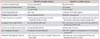
The changes in RECIST version 1.1 compared to version 1.0 are showed on Table 1. RECIST version 1.1's changed features are five categories: number of target lesions, assessment of pathologic lymph node, clarification of disease progression, clarification or baseline documentation of unequivocal progression of non-target lesions, inclusion of 18F-FDG-PET in the detection of new lesions11. Response criteria is divided into target and non-target lesion, and each evaluation are more specific than before. Revised RECIST criteria suggested standardized framework that can evaluate and analysis efficacy of cancer treatment. However, there are some limitations that evaluated certain organs (e.g., bone or liver) and some therapeutic options12. In addition, in response criteria, ideas of “30% decrease for a response” and “20% increase for a progression” are selected arbitrarily without enough evidence. And, the idea was applied to patient outcome such as overall survival which is problematic. For example, anti–vascular endothelial growth factor (VEGF), target therapeutic agent, decreased naïve tumor's size change which is lower than 30%, but significantly increased patient survival. In addition, there is some variation between patient outcome and response evaluation that became large sized scar to patients having image-guided therapy such as radiofrequency ablation or chemoembolization13.
In 2011, Lee et al.14 reported that lung cancer is generally measured on lung window from chest CT and included ground glass opacity (GGO) and solid components in part-solid lung cancer. However, GGO within part-solid lung cancer generally does not vary profoundly with cancer chemotherapy. Size change in only solid component of part-solid peripheral lung cancer, may be more accurate reflection of the actual tumor response to cancer for chemotherapy. Therefore, the article implied argument of RECIST measurement and RECIST solid measurement should be classified and applied to response criteria. In addition to patients with non-small cell lung cancer, lung image after chemotherapy show internal cavity formation due to necrosis of tumor. Cavitation within tumor caused by hampered angiogenesis, should be treated with platinum based chemotherapy and VGEF receptor blocker inhibitor for better response recoverd from damaged angiogenesis. Tumor necrosis may constitute a type of tumor response, but RECIST does not reflect this alternation15. In 2007, Choi et al.16 reported that lung cancer patients whom had chemotherapy had no alternation in tumor diameter evaluated from contrast chest CT, heterogeneously decreased attenuation. These alternation effected patient's survival rate. Therefore, alternation in attenuation implied tumor has shrunk and internal cavity formation, suggesting tumor necrosis. At this point, Choi response criteria was proposed. This criteria included that tumor attenuation which is Hounsfield Unit alternation of tumor attenuation.

Beyond RECIST Version 1.1

Evaluation of treatment response of solid tumor applied tumor size based RECIST, is widely applied and well accepted. However, as mentioned before, in new therapeutic modalities such as angiogenesis inhibitors and anti-vascular therapies, without size change, alternation such as attenuation, necrosis and cavitation, could be induced, underestimate could be a result in case of RECIST17. For example, using sorafenib and bevacizumab as metastatic renal cell cancer, clinicians could evaluate the disease as progressive state based on RECIST criteria: however, patient's condition was in of significantly approved and increased in progression-free survival18. After applying imatinib in gastrointestinal stromal tumor patient, clinician could observe significant alternation in standardized uptake values using by 18F-FDG-PET. In addition, clinicians could observe anatomical change by using CT several weeks later. There are limitations using CT which is bi-dimensional evaluative tool as the only evaluative method192021. Recently, many studies introduced the idea of “changes can precede volume changes” and insisted metabolic changes could be detected by different molecular and functional imaging techniques.
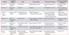
In 2016, there was a session of adjustment of functional or mixed functional and anatomical tumor response assessment at European Congression of Radiology (ECR) using newly introduced imaging modalities (Table 2). This meeting insisted response evaluation using new metabolic or functional imaging techniques, still acquired evaluation's end point for treatment efficacy as new targeted agents turned into main stream of cancer treatment even if it is in phase I or II trials. Also, these imaging modalities have idealistic merits: non-invasive, validated, and reproducible. Therefore, those imaging modalities introduced as new paradigm for tumor response evaluation222324252627282930.
With development of metabolic, and functional imaging modalities for evaluating target therapy's effect and efficacy, early change could be detected which leads productive selection for treatment strategy and prevents unnecessary long treatment course and adverse events. Thus patient's quality of life and cost effect possibly affected.

Immunotherapy Response Evaluation Methods

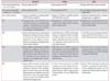
In cytotoxic agents, early increase in tumor growth and/or appearance of new lesions analyzed as progressive disease which means treatment failure. However, there need different comprehension for noncytotoxic agent which is immunotherapeutic agent. From mid 2000s, there had been active researches on immunotherapeutic agent and response evaluation. Table 3 is arranged immunotherapy methods that approached to lung cancer and malignant mesothelioma, up to date31. There were four kinds of response from melanoma patients whom had ipilimumab: two conventional response, response after tumor burden increase, and response in the presence of new lesion. Last two responses are was PD aspect of conventional response criteria but patients actually reacted to PR or stable disease. Delayed action mechanism of immunotherapy progresses T-cell expansion driving in first then T cell could expand which appears as tumor burden, or make cell edema from infiltration with tumor cell which could lead to radiography misinterpretation (a version of the “tumor flare reaction”)32.
To supplement this error, about 200 oncologists, immunotherapiests, and regulatory experts shared experiences of immunotherapeutic agents of cancer patients in 2004 to 2005, and concluded with following: the appearance of measurable anti-tumor activity may take longer for immune therapies than for cytotoxic therapies; responses to immune therapies may occur after conventional PD; discontinuation of immune therapy may not be appropriate in some cases, unless PD is confirmed (as is usually done for response); allowance for “clinical insignificant” PD (e.g., small new lesions in the presence of other responsive lesions) is recommended; and durable stable disease may represent anti-tumor activity. With this basis of conclusion, novel activity criteria for immunotherapeutic agents were introduced at Clinical Cancer Research in 2009. Comparing with conventional WHO criteria, its significant features are defined as following: new measurable lesion defined as incorporated into tumor burden, new non-measurable lesion do not define progression, sum of measurements defined as sum of uni-dimensional measurements of all target lesion and any new lesion. In immune related response criteria, immune related progressive disease defined as increase in tumor volume ≥20% from nadir, immune related stable disease as not meeting criteria for CR or PR, immune related partial response defined as decrease in tumor volume ≥30% relative to baseline, and immune related complete response defined as complete disappearance of all lesions and new measurable lesions. New lesion's definition was altered as presence of new lesions alone does not define progression and measurement of new lesions included in sum of measurement was included1533. Many retrospective studies applied new guideline, were proceeded until now, and in 2014, immune related RECIST was suggested at European Society for Medical Oncology. Thus, future prospective randomized study requires supplements and fixation by having more clinical trials, and researches on survival.

Conclusion

Even with the rapid development and variation of cancer therapeutic agents and imaging modalities, evaluation of chemotherapy is not sufficient. Therefore, evaluating new treatment options and imaging modalities applying WHO, RECIST version 1.0, or RECIST version 1.1, the evaluation end point (progression-free survival or overall survival) does not reflects satisfactorily. In addition, for global implementation of such novel treatment evaluation methods, multicenter or multi-institution research should not use original response criteria that could cause limitations for reproducibility.
Before appearance of new response criteria, many studies and discussion had conclusion of subjection toward original criteria and development of it. Thus, researchers, clinicians, and experts should have further studies and discussions for new response evaluating method appearance by acknowledge necessity of new method and developing new treatment and imaging modalities.

Figures and Tables

Table 1

Key features comparison of RECIST 1.0 and RECIST 1.1

trd-80-136-i001
RECIST 1.0 (2000 criteria) RECIST 1.1 (2009 criteria)
Assessment lymph node Not recommended Recommended
Assessment of tumor burden Five targets Ten targets
Two per organ Five per organ
Measuring lymph node Short axis Long axis as for other organs
Finding of a new lesion Not specifically defined Should be unequivocal
Patient response to treatment Initial response to treatment must be confirmed within 4 weeks Need not be confirmed in randomized trials when the primary end point is disease progression
Imaging of non-target lesions Not specifically addressed Not necessary at every protocol-specified for declaration of PR or SD
Lesions too small to measurement Not specifically defined Default value of 5 mm
New imaging techniques No specific recommendation FDG-PET applied to incorporate as CT scanning assessment
Definition of progressive disease >20% increase in sum of longest diameters form nadir >20% increase in sum of diameters from nadir, absolute increase of >5 mm

RECIST: Response Evaluation Criteria In Solid Tumor; PR: partial response; SD: stable disease; FDG-PET: fluorodeoxyglucose–positron emission tomography; CT: computed tomography.

Table 2

Beyond RECIST 1.1: new response criteria developed by clinical trials and new imaging modalities

trd-80-136-i002
Authors Published journal Name Clinical trials Assessment changes Alternative imaging modalities
Choi et al.16 J Clin Oncol (2007) Choi criteria Target therapy for GIST Size and attenuation at CT Perfusion CT, dual energy CT
Wahl et al.34 J Nucl Med (2009) PERCIST FDG-PET FDG uptake (SUVmax) PET (metabolism, oxygenation, other tracers)
Lencioni and Llovet35 Semin Liver Dis (2010) Lencioni criteria or modified RECIST Ablation therapy for hepatocellular carcinoma Size and arterial-phase Based enhancement at CT, MRI, or CEUS (contrast enhanced US) Perfusion MRI, diffusion-weighted MRI, perfusion US
Smith et al.36 AJR Am J Roentgenol (2010) MASS criteria Target therapy for metastatic renal carcinoma Size and attenuation at CT Perfusion CT, dual energy CT
Lee et al.14 Lung Cancer (2011) New response criteria (NRC) Target therapy for NSCLC Size and attenuation (solid portion) at CT Perfusion CT, dual energy CT
Chung et al.37 AJR Am J Roentgenol (2012) Modified CT criteria Chemotherapy for colorectal cancer liver metastasis Size and attenuation at CT Perfusion CT, dual energy CT
Nanni et al.38 Eur J Nucl Med Mol Imaging (2016) Italian Myeloma criteria for PET Use (IMPeTUs) Chemotherapy for multiple myeloma FDG uptake (baseline, induction, and end of treatment) FDG-PET/CT

RECIST: Response Evaluation Criteria In Solid Tumor; GIST: gastrointestinal stromal tumor; CT: computed tomography; PERCIST: Positron Emission Tomography Response Criteria in Solid Tumors; FDG-PET: fluorodeoxyglucose–positron emission tomography; SUVmax: maximum standardized uptake value; MRI: magnetic resonance imaging; CEUS: contrast-enhanced ultrasonography; US: ultrasonography; MASS: Morphology, Attenuation, Size, and Structure; NSCLC: non-small cell lung carcinoma.

Table 3

Comparison between WHO, RECIST version 1.1, and irRC criteria

trd-80-136-i003
RECIST WHO irRC
New measurable lesion (i.e., ≥5×5 mm) Always represent PD Always represent PD Incorporated into tumour burden
New non-measurable lesion (i.e., <5×5 mm) Always represent PD Always represent PD Do not define progression (but preclude irRC)
Non-index lesions Changes contribute to defining BOR of CR, PR, SD, and PD Changes contribute to defining BOR of CR, PR, SD, and PD Contribute to defining irRC (complete disappearance required)
CR Disappearance of all lesions in one observation in randomized studies; confirmation is needed for non-randomize d studies according to study protocol Disappearance of all lesions in two consecutive observations not less than 4 weeks apart Disappearance of all lesions in two consecutive observations not less than 4 weeks apart
PR At least a 30% decrease in the sum of diameters of target lesions, taking as reference the baseline sum diameters, in the absence of new lesions or unequivocal progression of non-index lesions A ≥50% decrease in SPD of all index lesions compared with baseline in observations, at least 4 weeks apart, in the absence of new lesions or unequivocal progression of non-index lesions A ≥50% decrease in tumour burden compared with baseline in two observations at least 4 weeks apart
SD Neither sufficient shrinkage to qualify for PR nor sufficient increase to qualify for PD, taking as reference the smallest sum diameters, in the absence of new lesions or unequivocal progression of non-index lesions A 50% decrease in SPD compared with baseline cannot be established, nor 25% increase compared with nadir, in absence of new lesions or unequivocal progression of non-index lesions A 50% decrease in tumour burden compared with baseline cannot be established, nor 25% increase compared with nadir
PD At least 20% increase in the sum of diameters of target lesions, taking as reference the smallest sum on study; the sum must also demonstrate an absolute increase of at least 5 mm; the appearance of one or more new lesions is also considered progression A t le as t 25% incr e ase in S P D compare d with nadir and/or unequivocal progression of non-index lesions and/or appearance of new lesions (at any single time point) At least 25% increase in tumour burden compared with nadir (at any single time point) in two consecutive observations at least 4 weeks apart

Adopted from Ades F and Yamaguchi N. Ecancermedicalscience 2015;9:604, according to the Creative Commons license31.

WHO: World Health Organisation; RECIST: Response Evaluation Criteria In Solid Tumor; irRC: immune-related response criteria; PD: progressive disease; BOR: best overall response; CR: complete response; PR: partial response; SD: stable disease; SPD: sum of the product of the greatest diameters.

Acknowledgments

This study was supported by grant of Wonkwang University in 2016.

Notes

Conflicts of Interest No potential conflict of interest relevant to this article was reported.

References

1. Miller AB, Hoogstraten B, Staquet M, Winkler A. Reporting results of cancer treatment. Cancer. 1981; 47:207–214.
2. Park JO, Lee SI, Song SY, Kim K, Kim WS, Jung CW, et al. Measuring response in solid tumors: comparison of RECIST and WHO response criteria. Jpn J Clin Oncol. 2003; 33:533–537.
3. Therasse P. Measuring the clinical response: what does it mean? Eur J Cancer. 2002; 38:1817–1823.
4. James K, Eisenhauer E, Christian M, Terenziani M, Vena D, Muldal A, et al. Measuring response in solid tumors: unidimensional versus bidimensional measurement. J Natl Cancer Inst. 1999; 91:523–528.
5. Prasad SR, Saini S, Sumner JE, Hahn PF, Sahani D, Boland GW. Radiological measurement of breast cancer metastases to lung and liver: comparison between WHO (bidimensional) and RECIST (unidimensional) guidelines. J Comput Assist Tomogr. 2003; 27:380–384.
6. Padhani AR, Husband JE. Commentary: are current tumour response criteria relevant for the 21st century? Br J Radiol. 2000; 73:1031–1033.
7. Eisenhauer EA, Therasse P, Bogaerts J, Schwartz LH, Sargent D, Ford R, et al. New response evaluation criteria in solid tumours: revised RECIST guideline (version 1.1). Eur J Cancer. 2009; 45:228–247.
8. Nishino M, Jackman DM, Hatabu H, Yeap BY, Cioffredi LA, Yap JT, et al. New Response Evaluation Criteria in Solid Tumors (RECIST) guidelines for advanced non-small cell lung cancer: comparison with original RECIST and impact on assessment of tumor response to targeted therapy. AJR Am J Roentgenol. 2010; 195:W221–W228.
9. Therasse P, Eisenhauer EA, Verweij J. RECIST revisited: a review of validation studies on tumour assessment. Eur J Cancer. 2006; 42:1031–1039.
10. van Persijn van Meerten EL, Gelderblom H, Bloem JL. RECIST revised: implications for the radiologist. A review article on the modified RECIST guideline. Eur Radiol. 2010; 20:1456–1467.
11. Bogaerts J, Ford R, Sargent D, Schwartz LH, Rubinstein L, Lacombe D, et al. Individual patient data analysis to assess modifications to the RECIST criteria. Eur J Cancer. 2009; 45:248–260.
12. Trillet-Lenoir V, Freyer G, Kaemmerlen P, Fond A, Pellet O, Lombard-Bohas C, et al. Assessment of tumour response to chemotherapy for metastatic colorectal cancer: accuracy of the RECIST criteria. Br J Radiol. 2002; 75:903–908.
13. Rasmussen F, Madsen HH. Imaging follow-up of RF ablation of lung tumours. Cancer Imaging. 2011; 11 Spec No A:S123–S128.
14. Lee HY, Lee KS, Ahn MJ, Hwang HS, Lee JW, Park K, et al. New CT response criteria in non-small cell lung cancer: proposal and application in EGFR tyrosine kinase inhibitor therapy. Lung Cancer. 2011; 73:63–69.
15. Hoos A, Eggermont AM, Janetzki S, Hodi FS, Ibrahim R, Anderson A, et al. Improved endpoints for cancer immunotherapy trials. J Natl Cancer Inst. 2010; 102:1388–1397.
16. Benjamin RS, Choi H, Macapinlac HA, Burgess MA, Patel SR, Chen LL, et al. We should desist using RECIST, at least in GIST. J Clin Oncol. 2007; 25:1760–1764.
17. Nishino M, Jagannathan JP, Krajewski KM, O'Regan K, Hatabu H, Shapiro G, et al. Personalized tumor response assessment in the era of molecular medicine: cancer-specific and therapy-specific response criteria to complement pitfalls of RECIST. AJR Am J Roentgenol. 2012; 198:737–745.
18. Hutson TE. Targeted therapies for the treatment of metastatic renal cell carcinoma: clinical evidence. Oncologist. 2011; 16:Suppl 2. 14–22.
19. Yoshida S, Miyata Y, Ohtsu A, Boku N, Shirao K, Shimada Y. Significance of and problems in adopting Response Evaluation Criteria in Solid Tumor RECIST for assessing anticancer effects of advanced gastric cancer. Gastric Cancer. 2000; 3:128–133.
20. Park JY, Jang SH. Epidemiology of lung cancer in Korea: recent trends. Tuberc Respir Dis (Seoul). 2016; 79:58–69.
21. Tuma RS. New response criteria proposed for immunotherapies. J Natl Cancer Inst. 2008; 100:1280–1281.
22. Desar IM, van Herpen CM, van Laarhoven HW, Barentsz JO, Oyen WJ, van der Graaf WT. Beyond RECIST: molecular and functional imaging techniques for evaluation of response to targeted therapy. Cancer Treat Rev. 2009; 35:309–321.
23. Fournier L, Ammari S, Thiam R, Cuenod CA. Imaging criteria for assessing tumour response: RECIST, mRECIST, Cheson. Diagn Interv Imaging. 2014; 95:689–703.
24. Erasmus JJ, Gladish GW, Broemeling L, Sabloff BS, Truong MT, Herbst RS, et al. Interobserver and intraobserver variability in measurement of non-small-cell carcinoma lung lesions: implications for assessment of tumor response. J Clin Oncol. 2003; 21:2574–2582.
25. Kauczor HU, Zechmann C, Stieltjes B, Weber MA. Functional magnetic resonance imaging for defining the biological target volume. Cancer Imaging. 2006; 6:51–55.
26. McLarty K, Reilly RM. Molecular imaging as a tool for personalized and targeted anticancer therapy. Clin Pharmacol Ther. 2007; 81:420–424.
27. Rohren EM, Turkington TG, Coleman RE. Clinical applications of PET in oncology. Radiology. 2004; 231:305–332.
28. Glade Bender J, Cooney EM, Kandel JJ, Yamashiro DJ. Vascular remodeling and clinical resistance to antiangiogenic cancer therapy. Drug Resist Updat. 2004; 7:289–300.
29. Meijerink MR, van Cruijsen H, Hoekman K, Kater M, van Schaik C, van Waesberghe JH, et al. The use of perfusion CT for the evaluation of therapy combining AZD2171 with gefitinib in cancer patients. Eur Radiol. 2007; 17:1700–1713.
30. Kawada K, Murakami K, Sato T, Kojima Y, Ebi H, Mukai H, et al. Prospective study of positron emission tomography for evaluation of the activity of lapatinib, a dual inhibitor of the ErbB1 and ErbB2 tyrosine kinases, in patients with advanced tumors. Jpn J Clin Oncol. 2007; 37:44–48.
31. Ades F, Yamaguchi N. WHO, RECIST, and immune-related response criteria: is it time to revisit pembrolizumab results? Ecancermedicalscience. 2015; 9:604.
32. Wolchok JD, Hoos A, O'Day S, Weber JS, Hamid O, Lebbe C, et al. Guidelines for the evaluation of immune therapy activity in solid tumors: immune-related response criteria. Clin Cancer Res. 2009; 15:7412–7420.
33. Wolchok JD, Neyns B, Linette G, Negrier S, Lutzky J, Thomas L, et al. Ipilimumab monotherapy in patients with pretreated advanced melanoma: a randomised, double-blind, multicentre, phase 2, dose-ranging study. Lancet Oncol. 2010; 11:155–164.
34. Wahl RL, Jacene H, Kasamon Y, Lodge MA. From RECIST to PERCIST: evolving considerations for PET response criteria in solid tumors. J Nucl Med. 2009; 50:Suppl 1. 122S–150S.
35. Lencioni R, Llovet JM. Modified RECIST (mRECIST) assessment for hepatocellular carcinoma. Semin Liver Dis. 2010; 30:52–60.
36. Smith AD, Shah SN, Rini BI, Lieber ML, Remer EM. Morphology, Attenuation, Size, and Structure (MASS) criteria: assessing response and predicting clinical outcome in metastatic renal cell carcinoma on antiangiogenic targeted therapy. AJR Am J Roentgenol. 2010; 194:1470–1478.
37. Chung WS, Park MS, Shin SJ, Baek SE, Kim YE, Choi JY, et al. Response evaluation in patients with colorectal liver metastases: RECIST version 1.1 versus modified CT criteria. AJR Am J Roentgenol. 2012; 199:809–815.
38. Nanni C, Zamagni E, Versari A, Chauvie S, Bianchi A, Rensi M, et al. Image interpretation criteria for FDG PET/CT in multiple myeloma: a new proposal from an Italian expert panel. IMPeTUs (Italian Myeloma criteria for PET USe). Eur J Nucl Med Mol Imaging. 2016; 43:414–421.
TOOLS
Similar articles