

 PDF
PDF ePub
ePub Citation
 Print
Print


Introduction
An endobronchial blood clot is an unsusal, albeit not rare cause of airway obstruction1,2,3. In spontaneously breathing patients, the clinical features of endobronchial blood clots may resolve rapidly when blood clots are coughed up. Endobronchial blood clot formation is not always preceded by hemoptysis prior to the development of sudden airway obstruction particularly in patients on mechanical ventilation4. We report a case of airway obstruction due to endobronchial blood clots during mechanical ventilation. The clots were removed using bronchoscopic cryotherapy at bedside of intensive care unit (ICU).
Case Report
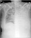
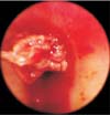
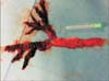
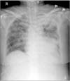
A 66-year-old female with endometrial cancer who had undergone chemotherapy with doxorubicin and cisplatin 8 days previously, presented to the hospital with neutropenic fever and pancytopenia. She was admitted to the hospital and treated with broad-spectrum antibiotics. Three days after admission, she developed hypoxic respiratory failure requiring intubation (endotracheal tube diameter, 8.0 mm) for acute pulmonary edema and septic shock. During mechanical ventilation, the minute ventilation suddenly dropped to significantly low levels and mildly blood-tinged material was obtained when suctioning the endotracheal tube. Chest radiography revealed no change from previous radiography. Next morning; 12 hours later, chest radiography showed a completely opacified left hemithorax (Figure 1). On auscultation, there were no breath sounds over the left chest. The patient's platelets were 38,000/µL. Flexible bronchoscopy (5.9 mm diameter, 2.8 mm channel diameter; BF-1T260; Olympus, Tokyo, Japan) at bedside of ICU revealed a large blood clot obstructing the proximal left main bronchus (Figure 2). Because of the friable structure of the clot, bronchoscopic lavage and forceps extraction were unsuccessful. After repeated attempts to remove the blood clot over one hour, a flexible cryoprobe (2.4 mm diameter; ERBE, Tubingen, Germany) was applied via flexible bronchoscopy to the blood clot and frozen for 10 seconds. The blood clot firmly attached to the probe and was successfully removed in four large pieces (Figures 3, 4). A follow-up chest X-ray revealed significant improvement in aeration of the left lung (Figure 5). Fifteen minutes were required to remove the blood clots using bronchoscopic cryotherapy. No further obstructive events occurred. A repeat bronchoscopic evaluation 2 weeks later showed no evidence of the clot and all of the airways were patent. She recovered from septic shock and discharged from hospital on the 24th day.
Discussion
Acute airway obstruction due to the presence of blood clots occurs in a variety of clinical conditions, including complications of respiratory disease, such as tuberculosis, bronchiectasis, bronchial carcinoma, and pulmonary arteriovenous malformations, as well as other causes, such as anticoagulation or pancytopenia1,2,3,4,5,6. These conditions may result in lifethreatening impairment of ventilation. The initial efforts to remove an endobronchial clot should involve flexible bronchoscopic evaluation with saline irrigation and suction7. Forceps extraction through the working channel or removal of the clot with the forceps and bronchoscope together may be necessary. If all of the aforementioned methods fail, the next step is rigid bronchoscopy. Rigid bronchoscopy provides greater advantages in terms of direct suctioning and forceps extraction with large rigid bore suction catheters and forceps. However, patients undergoing rigid bronchoscopy need to be extubated before the procedure. Although mechanical ventilation can be performed through a side-port channel, the previously impaired ventilator status may be worse. Furthermore this technique requires the expertise of specialists. Recently, alternative methods for removing endobronchial blood clots have been introduced, such as a Fogarty arterial embolectomy catheter or application of thrombolytics via a flexible bronchoscopy8,9. These methods are only recommended when severe respiratory compromise is present and other alternatives have failed. Bronchoscopic cryotherapy has been used for the biopsy of airway pathology and the ablation of endobronchial lesions, such as a malignant masses or benign tracheal stenosis10,11,12. Owing to its freezing characteristics, bronchoscopic cryotherapy can also be used to remove foreign bodies that contain sufficient water, a procedure referred to as cryoextraction13. During cryoextraction, tissue or foreign bodies are frozen to the tip of the probe and removed by pulling on the probe together with the bronchoscopy. The development of a thin and flexible cryoprobe has allowed bronchoscopic cryotherapy without the risks associated with general anesthesia and rigid bronchoscopy14.
In this case of our patient, we removed blood clots that induced ventilation dysfunction without extubation and general anesthesia, which are both required for rigid bronchoscopy. Furthermore, we conducted bronchoscopic cryotherapy at bedside in ICU. It is foreseeable that removal of blood clots using rigid bronchoscopy may lead to a prolonged procedure time due to the removal of only small pieces of the clot with each forceps bite. In contrast, the bronchoscopic cryotherapy technique allows en-bloc removal of the blood clot. An additional advantage of this technique is that the learning curve to utilize the cryotherapy probe is quite short, and does not require the more extended training needed to master rigid bronchoscopy; something which is not available in many institutions.
Bronchoscopic cryotherapy usually applied after blood clots formation. Previous study described the endoscopic use of cryotherapy to treat actively bleeding diffuse mucosal vascular lesions in gastrointestinal tract15. Argon plasma coagulation, laser, and other thermal modalities have the potential for deep tissue injury with ulceration and bleeding. Cryotherapy resulted in only superficial tissue damage that usually was completely healed by 3 months in the study. Bronchoscopic cryotherapy could be alternative modality to treat not only blood clots but also active bleeding mucosal lesions in bronchus. One concern about removal endobronchial blood clots using bronchoscopic cryotherapy is that a large blood clot might be difficult to extract through the small sized endotracheal tube.
To our knowledge, this is the first case to report the utilization of bronchoscopic cryotherapy to remove endobronchial blood clots at bed side of ICU. It highlights the utility of cryotherapy probes through flexible bronchoscopy to remove any object, with sufficient water content at bed side of ICU. We conclude that these properties of the procedure assisted us in the safe and easy removal of endobronchial blood clots at the ICU bedside, and thus, avoided the use of more invasive approaches such as rigid bronchoscopy.

 XML Download