

 PDF
PDF ePub
ePub Citation
 Print
Print


Abstract
In 2005, a group of mycolic acid-containing bacteria was characterized as belonging to a novel genus, Segniliparus with species Segniliparus rugosus and S. rotundus. We report a case of the S. rugosus isolated from a 54-year-old woman with radiologic features mimicking that of non-tuberculous mycobacteriosis (NTM). When the patient first visited our hospital, an acid-fast bacteria (AFB) smear tested positive and Mycobacterium tuberculosis polymerase chain reaction (TB PCR) was negative in the bronchoalveolar lavage sample. After 2 months, the growing colonies were reported as NTM, but could not be identified because they had died. One year after the initial visit, induced sputum samples showed the same results, positive AFB smear and negative TB PCR. At this point, the growing colonies were identified as S. rugosus. Therefore, we should consider Segniliparus genus as a differential diagnosis for AFB in respiratory specimens in addition to the genus Mycobacterium.
Figures and Tables
Figure 1
Chest X-ray images. (A) Chest X-ray taken 7 months before the first OPD visit showed parenchymal infiltration in the left middle lung field. (B) Chest X-ray taken during the first visit showed persistent parenchymal infiltration in left middle lung field. OPD: outpatient department.
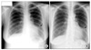
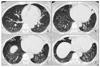
Figure 2
Chest HRCT images taken during the first visit revealed small nodules and branching centrilobular opacities in the left lingular segment (A), RML (B, C) and consolidation containing air bronchogram in the left lingular segment (B, C) and RML (D). HRCT: high resolution computed tomography; RML: right middle lobe.
References
1. Butler WR, Floyd MM, Brown JM, Toney SR, Daneshvar MI, Cooksey RC, et al. Novel mycolic acid-containing bacteria in the family Segniliparaceae fam. nov., including the genus Segniliparus gen. nov., with descriptions of Segniliparus rotundus sp. nov. and Segniliparus rugosus sp. nov. Int J Syst Evol Microbiol. 2005. 55:1615–1624.
2. Butler WR, Sheils CA, Brown-Elliott BA, Charles N, Colin AA, Gant MJ, et al. First isolations of Segniliparus rugosus from patients with cystic fibrosis. J Clin Microbiol. 2007. 45:3449–3452.
3. Butler WR, Floyd MM, Silcox V, Cage G, Desmond E, Duffey PS, et al. Standardized method for HPLC identification of Mycobacteria. 1996. Atlanta, GA: CDC, US Department of Health and Human Services.
4. Floyd MM, Gross WM, Bonato DA, Silcox VA, Smithwick RW, Metchock B, et al. Mycobacterium kubicae sp. nov., a slowly growing, scotochromogenic Mycobacterium. Int J Syst Evol Microbiol. 2000. 50:1811–1816.
5. Griffith DE, Aksamit T, Brown-Elliott BA, Catanzaro A, Daley C, Gordin F, et al. An official ATS/IDSA statement: diagnosis, treatment, and prevention of nontuberculous mycobacterial diseases. Am J Respir Crit Care Med. 2007. 175:367–416.
6. Hansen T, Van-Kerckhof J, Jelfs P, Wainwright C, Ryan P, Coulter C. Segniliparus rugosus infection, Australia. Emerg Infect Dis. 2009. 15:611–613.
 XML Download