

 PDF
PDF ePub
ePub Citation
 Print
Print


Introduction
If there is a communication between alveoli and pleura, the air in the pleural space is observed on chest radiography. It is also seen in the cases with direct communication between atmosphere and pleural space, or the presence of gas-producing organisms in the pleural space. It includes pneumothorax and bronchopleural fistula. They are caused by idiopathic and may also occur after traumatic, invasive interventions or pulmonary resection. The goal of the treatment is to remove the air present in the pleural space and to prevent secondary recurrence. Chest tube drainage is the most commonly used treatment method. If thoracoscopy is selected, transient chest tube drainage may also be required.
During the treatment of chest tube drainage, air leak may be persistent from the tube. It is mainly attributable to the presence of bronchopleural fistula or air leak through the bronchiole, which requires medical or surgical treatment. If a surgical treatment is difficult to perform due to underlying illness, medical treatments should be selected. The flexible bronchoscopy has allowed us to close the fistula using tissue glues, fibrin glues, sealants or sclerong agents1-4.
We hypothesized that there were certain communications between alveoli (or bronchioles) and pleural space in patients with persistent air leak from the chest tube. Thus, we instilled ethanolamine solution into the target bronchus bronchoscopically and evaluate the clinical outcomes after this therapy. This procedure may reduce medical costs and the duration of hospitalization.
Materials and Methods
1. Subjects
During chest tube drainage for pneumothorax or empyema, the patients who had persistent air leak and incomplete expansion of the lung due to the air in the pleural space were included. All patients provided written informed consent, and they agreed to be treated with bronchoscopic ethanolamine injection. But, in the cases of bronchopleural fistula formation after pulmonary resection, they were excluded in this study because surgical correction needed to be inevitable.
2. Methods
For bronchoscopic preparations, meperidine 25~50 mg and atropine sulfate 0.5 mg were injected intramuscularly 15~30 minutes before the bronchoscopy. If necessary, midazolam 1.0~2.0 mg was injected intravenously for further sedation. Before beginning the procedure, the patients were nebulized by 2% lidocaine hydrochloride 10 mL for the local anesthetic effect of the oropharynx.
Bronchoscopy was conducted to identify the presence of endobronchial lesions by conventional methods. If bronchopleural fistula was seen grossly during bronchoscopy, 5% ethanolamine oleate 1.0 mL was instilled to the surrounding mucosa of the orifice repeatedly via the injection needle inserted through the bronchoscopic biopsy channel.
If no lesion was found, the bronchoscope was fixed to the lobar bronchus that was most likely to have the air leak sites. In consideration of radiologic and postoperative findings, we select segmental or subsegmental bronchus and wedge the target bronchus with the bronchoscope. Then, the patients were asked to cough to see if air bubbles from the chest tube were significantly reduced or halted. If so, it was concluded that air leak occurred through the segmental (or subsegmental) bronchus. The injection catheter was placed in the target bronchus for passing through bronchoscopic channel and catheter tip out from the distal end. The aliquots of 5% ethanolamine oleate 1.0 mL were injected through the catheter repeatedly. If the patients complained of discomfort or pain during the procedure, the therapy was discontinued.
If air leak from the chest tube was halted by ethanolamine injection therapy and the chest tube was removed from the patients after the complete expansion of the lung, the treatment was considered to be successful. If other treatments such as surgery were required, however, the treatment was considered to be unsuccessful.
Results
1. Basal clinical characteristics
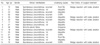
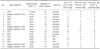
A total of 15 patients were enrolled in the study. They were all male patients aged 16~75 years. The clinical manifestations of the patients consisted of 14 cases (primary 7 cases, recurrent 7 cases) of pneumothorax and one case of hydropneumothorax. Underlying lung diseases included 9 cases of idiopathic (60%), 3 cases of chronic obstructive pulmonary disease (COPD, 20%), 2 cases of tuberculosis or past history of tuberculosis (13.3%), and once case of empyema associated with bronchopleural fistula (6.7%). Among the seven patients with recurrent pneumothorax, five patients had a previous history of surgical treatments (Table 1).
Five patients had incomplete lung expansions due to persistent air leak despite a surgical treatment for apical bleb during the admission (Table 2). A chest computed tomography (CT) was done in 13 patients, among whom blebs or bullae were observed in 12 patients and pleural effusion was observed in one patient.
2. Treatment outcomes
Ethanolamine injection therapy was conducted 1~3 times: once in 12 patients (80%), twice in two patients (13.3%), and three times in one patient (6.7%). The total injection amount of ethanolamine was between 5 mL (minimum) and 21 mL (maximum). An Ethanolamine injection therapy was initiated 1~18 days (median, 3.5 days) after the initiation of chest tube drainage. In the cases of pneumothorax with surgical therapy (5 cases), an ethanolamine was injected 0~8 days (median, 2 days) after the operation. Successful outcomes were shown in 12 patients (80%). Meanwhile, the treatment was considered unsuccessful in the three patients (20%), followed by additional surgery in two patients and an autologous blood patch pleurodesis in one patient. In the cases of successful treatment, 1~11 days (median, 3 days) were needed for the removal of the chest tube and 1~13 days (median, 3 days) were taken to discharge from the hospital (Table 2).
3. Complication
After an ethanolamine injection therapy, the patients experienced any of the following adverse reactions: fever (4 cases, 26.7%), chest pain (2 cases, 13.3%), fever and chest pain (4 cases, 26.7%). Radiographic findings after ethanolamine therapy showed no significant changes (6 cases, 40%), patchy pulmonary infiltrations (8 cases, 83.3%), and hydropneumothorax (1 case, 6.7%). All of them showed transient courses and were gradually resolved clinically and radiographically as time passed by.
Discussion
An ethanolamine injection therapy was initially introduced to control gastrointestinal bleeding5. It is also used for the extracting an intraluminal polypoid tumor that is protruding into the airway in respiratory area6. An ethanolamine injection therapy is effective in inducing rapid dehydration and scar formation of the tissue. These effects are also applied to the treatment of early gastric cancer as regional edema, necrosis and ulcer formation are anticipated with local injection of ethanolamine solution5,7. Histologically, fibroblast proliferation and tissue reconstruction by fibrous scar can be observed within three weeks2,7. The authors focused on the regional edema and chemical inflammatory reaction of an ethanolamine on tissues. Accordingly, this study was conducted to investigate the effect of ethanolamine injection therapy in patients with pneumothorax, particularly, those who were unable to remove the chest tube quickly due to persistent air leak from chest tube drainage.
An ethanolamine injection therapy was conducted in 15 patients (pneumothorax 14 cases, empyema one case caused by bronchopleural fistula). The catheter was fixed to the segmental bronchus that was suspected to have air leak, and the aliquots of 5% ethanolamine oleate 1.0 mL were slowly injected 5~21 times through the catheter. Because the rapid injection of high dose has been reported to cause deep ulcer and tissue toxicity, leading to necrosis in the tissue5,6, an ethanolamine solution was slowly injected at a low dose in this study. As a result, the chest tube was successfully removed from 12 patients (88%).
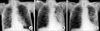
In Figure 1 (case 1), a recurrent large pneumothorax in left side occurred in a 75-year-old male patient who had been on the outpatient department for post-tuberculous bronchiectasis and emphysematous lung. Despite chest tube drainage, the lung was not expanded fully and a large amount of air leak via the chest tube was persistent until 7 days later. Thus, a surgical treatment was required. However, as his sputum acid-fast bacillus smear and post-bronchodilator forced expiratory volume in one second was shown to be positive and only 630 mL (27% of predicted value) respectively, he had a high risk for surgical treatment. Thus, the patient underwent bronchoscopic ethanolamine injection therapy total three times. Thereafter, air leak was halted and the lung was fully expanded, and so the chest tube was successfully removed. The patient was discharged from the hospital with anti-tuberculous medications (Figure 1). It has been known that air leak from spontaneous pneumothorax that occurs in the case of emphysema-like changes caused by tuberculous destroyed lung and COPD is resulted from the rupture of bulla8. Thus, air leak site or any abnormalities resulting probably into air leak should be corrected to prevent further recurrences. Currently, thoracoscopic treatment for emphysema-like changes (blebs or bullae) and pleurodesis have been commonly used9,10. In some cases, wedge resection with bullae placation is preferred depending on thoracic surgeons. If the air leak persist more than 4 days even in the first episode of spontaneous pneumothorax, a surgical treatment should be considered11. However, if the patient had a high risk of anesthesia or surgery due to underlying illness or poor pulmonary function, other therapeutic modalities should be alternatively selected, but there are no effective treatments until now.
Although the injection of sclerosing agents (tetracycline, minocyclin, doxycyclin or talc slurry) via the chest tube can be used, the recurrence rate of pneumothorax has been reported to be 15~25%12-14. Thus, an ethanolamine injection therapy can be considered as an alternative treatment option.
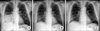
In Figure 2 (case 9), a 56-year-old male patient who had no specific underlying lung disease visited the hospital due to fever and chest pain. After right hydropneumothorax was observed on radiography, the patient immediately underwent chest tube drainage. As a bronchopleural fistula was suspected on a chest CT, bronchoscopy was conducted prior to the surgical treatment. As a result, a small fistula was observed in the orifice of right middle lobe bronchus, from which pus-like foamy secretion was discharged. The patient was diagnosed with empyema caused by a bronchopleural fistula, and the air leak from the chest tube was expected to be halted if this fistula was closed. Luckily, the air leak was halted after ethanolamine injection therapy, and subsequently, the chest tube was removed from the patient. Takaoka et al.15 reproted that local injection of an ethanolamine was effective in occluding central bronchopleural fistula with a diameter of 1 mm or less because the swollen mucosa epithelium due to regional edema induced the mechanical occlusion of the fistula. That is, if an ethanolamine is injected to the mucosa around the fistula orifice, increased blood flow, fibrin disintegration and tissue granulation are formed as the time went by, and 2 months later, connective tissue and fibrin were completely covered over with epithelial tissue2,5. Subsequently, the fistula was covered with fibrous scarring. However, in the case of fistula with a diameter of 3 mm or more, it is difficult to close because the patient would expectorate the fibrin plug by local injection of ethanolamine solution during coughing even if it closes the fistula initially. It has been reported that the mortality is high due to aspiration pneumonia, pyothorax, and sepsis if a surgical treatment is conducted in patients with bronchopleural fistula. Hollaus et al.16 reported that postoperative mortality was 31%. Therefore, in the treatment of bronchopleural fistula, a bronchoscopic treatment may be an alternative prior to the determination of surgical treatment.
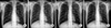
As bronchoscopic ethanolamine injection therapy can be conducted under local anesthesia, light anesthesia or analgesics, it can avert risks associated with general anesthesia. Small amounts of over-flowed ethanol induced coughing, chest discomfort, and fever, but no serious respiratory complications such as hypoxia occurred during ethanolamine injection therapy. As radiologic opacities were shown in 8 patients (53.3%), ethanolamine-induced pneumonia was suspected. However, they were spontaneously resolved within 2~3 days (case 10) (Figure 3B~D). This was likely to be attributable to the regional edema due to ethanolamine injection5,7.
In addition, ethanolamine injection therapy has costeffective benefits. If this treatment is successful, invasive procedures such as surgery are not required and in-hospital stays can also be reduced. In this study, 1~13 days (median, 3 days) were taken from the last injection of ethanolamine to hospital discharge in 12 patients.
In conclusion, an bronchoscopic ethanolamine injection therapy can avoid surgical treatments and reduce the duration of hospitalization in patients with persistent air leak from chest tube drainage and incomplete lung expansion. However, a further study is required to investigate the appropriate inclusion criteria for the treatment, treatment frequency, and safety. In this study, unsatisfactory results were obtained from three patients (20%) (Table 2). In the cases of idiopathic and spontaneous pneumothorax associated with post-tuberculous scar, lung expansion did not get fully even after two times of ethanolamine injection therapy, and a surgical treatment was required. In the patient with COPD, multiple bullae were seen on the chest CT and so, a surgical treatment was recommended for the prevention of recurrence instead of repeated ethanolamine therapy, but the patient wanted non-surgical therapy and received autologous blood patch pleurodesis to halt air leak17. It is unclear when ethanolamine injection therapy should be conducted in patients with persistent air leak from chest tube drainage. In this study, an ethanolamine injection therapy was first conducted on 1~18 days (median, 3.5 days) after introduction of chest tube drainage. It was initially planed to be conducted 4 days after chest tube drainage. However, it was conducted earlier when the patients showed persistent air leak and incomplete lung expansion despite chest tube drainage or thoracic surgeons needed the therapy after operation, whereas it was often delayed up to 10 days than the initial schedule in the cases of underlying illness, bronchopleural fistula and surgical candidates.
 XML Download