

 PDF
PDF ePub
ePub Citation
 Print
Print


Introduction
Mycobacterium terrae complex is known to comprise multiple species, including M. terrae, M. nonchromogenicum, and M. trivia1. M. terrae is ubiquitous in the environment and generally considered a nonpathogenic nontuberculous mycboacteria (NTM)1. The most common presentation of M. terrae infection is tenosynovitis of the upper extremities, often following trauma2,3. To date, few cases of NTM lung disease caused by M. terrae have been reported. Here, we report a very rare case of M. terrae lung disease associated with bronchiectasis in an immunocompetent patient.
Case Report
In March 2008, a 45-year-old woman was referred to our hospital due to chronic cough and sputum. The patient had been healthy until two months earlier, when cough and purulent sputum developed. She had a history of smoking (10 pack-years), but had stopped 10 years prior. The patient had pectus excavatum. Laboratory results were normal with the exception of elevated CA 19-9 (75.32 U/mL, normal range, 0~37 U/mL). Her height was 161 cm and body weight was 51 kg. A human immunodeficiency virus (HIV) antibody test was negative.
A computed tomography (CT) scan of the chest revealed bronchiectasis and bronchiolitis in both lungs (Figure 1A). The patient also had pectus excavatum in the anterior chest wall. There was no evidence of cystic fibrosis or other common causes of bronchiectasis. NTM were isolated from bronchoalveolar lavage fluid collected from both lungs and cultured in vitro.
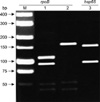
Polymerase chain reaction (PCR) restriction fragment length polymorphism analysis (PRA) was used to identify the particular mycobacterial species isolated from the patient. DNA was extracted from pure culture colonies of mycobacteria grown on 7H10-OADC agar plate and the genes rpoB and hsp65 were amplified by PCR. PCR product was digested using the restriction enzymes MspI and HindIII as described previously4,5. The pattern of the resulting DNA fragments was then compared to those kept in a database of known mycobacterial species. PRA of the rpoB gene resulted in fragment lengths of 110, 95, 55, and 45 bp using MspI and 175, 55, and 45 bp using HaeIII (Figure 2). These DNA fragment lengths matched the known restriction fragment patterns of the reference strain of M. terrae ATCC15755 reported in the previous studies5,6. The identity of etiological agent was also confirmed by sequencing analysis, revealing 99 and 100% sequence similarity with the rpoB gene (GenBank accession no. AF057488.1) and hsp65 gene (GenBank accession no. AF547879) of M. terrae ATCC15755, respectively.
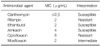
Drug susceptibility testing was performed using a broth microdilution method, as previously described for slow growing mycobacteria, such as M. kansasii, according to the guidelines of the Clinical and Laboratory Standards Institute (CLSI)7. The clinical isolate of M. terrae was susceptible to clarithromycin, ethambutol, and amikacin but resistant to ciprofloxacin and rifampin (Table 1).
The patient was treated with oral clarithromycin (1,000 mg/day), rifampin (600 mg/day), and ethambutol (800 mg/day) for 18 months. The treatment outcome was favorable; the patient's symptoms resolved completely, radiographic findings improved (Figure 1B), and negative conversion of sputum cultures was achieved and maintained after 12 months of antibiotic therapy (Figure 1B).
Discussion
Despite the prevailing view that M. terrae complex isolates are nonpathogenic, these organisms are occasionally identified in clinical specimens8. For example, in our institution, M. terrae complex made up 3% of 1,548 NTM clinical isolates during the 2-year period from 2002 to 20039. Nonetheless, no patient had clinically significant disease caused by M. terrae complex infection during that time.
NTM lung disease caused by M. terrae complex is rarely reported in the literature10-12, and there is no reported case in Korea. Furthermore, previous studies have had difficulty identifying the exact species of clinical isolates, as commercial DNA probes and high performance liquid chromatography (HPLC) are not suitable methods to distinguish between different member species of the M. terrae complex. A recent study analyzing 16S rRNA and hsp65 genes demonstrated genomic heterogeneity among M. terrae complex members13. Therefore, we were able to confirm that our patient had M. terrae infection using modern molecular techniques that enabled sequencing of the rpoB and hsp65 genes.
NTM lung disease has two distinct radiographic manifestations: an upper lobe fibrocavitary form and a nodular bronchiectatic form1. The nodular bronchiectatic form of the disease occurs predominantly in non-smoking middle-aged or elderly women who have no underlying lung disease. The radiographic features of the nodular bronchiectatic form include bronchiectasis and multiple nodules, which tend to be most severe in the lingular segment of the left lung and in the right middle lobe. These features are well characterized in M. avium complex lung disease, but have also been noted in NTM lung disease caused by other NTM species, such as M. abscessus complex14-16. In addition, women with the nodular bronchiectatic form of NTM lung disease also often have certain medical conditions, including pectus excavatum, scoliosis, and thin body habitus17. Our case study presented with typical clinical and radiographic features of the nodular bronchiectatic form of NTM lung disease.
The optimal antimicrobial therapy regimen for M. terrae infection has yet to be established. Some have suggested that drug treatment regimen for M. terrae infection should include macrolide, rifampin, and ethambutol2. Our patient received these three drugs and had substantial improvement without significant sequelae. Despite the rifampin resistant that the clinical isolates exhibited in vitro, it was included in the treatment regimen for our patient. It is unclear whether the combination of rifampin and ethambutol proved an additive, or synergistic, effect2.
In conclusion, M. terrae should be considered a possible etiologic pathogen of the nodular bronchiectatic form of NTM lung disease, despite the rare occurrence of M. terrae lung disease. Additionally, macrolide-containing antibiotic therapies may be effective in the treatment of M. terrae lung disease.

 XML Download