Abstract
Background
Lung abscess is necrosis of the pulmonary parenchyma caused by microbial infection. At present, clinical outcomes after treatment are good. However, the pulmonary parenchymal changes on the chest computed tomography (CT) after treatment are not well known. We studied the changes of pulmonary parenchyma on plane chest radiography and chest CT in patients with lung abscess following the administration of antibiotics.
Methods
We retrospectively reviewed 39 patients who had lung abscess with or without combined pneumonia from January 2006 to July 2010. We studied the therapeutic response in plane chest radiography of them at 1, 2, or more than 3 months following treatment. If any chest CT of them during the study period, we reviewed.
Results
Mean age of the patients was about 61.3±11.2. Mean duration of antibiotics administration was about 36.7±26.8 days. After 3 months of following plane chest radiography, 10 patients (36%) showed without residual sequelae among 28 patients. Findings from other patients showed decrease in densities (11 patients, 39%), fibrostreaky sequelae (4 patients, 14%) and bullae (3 patients, 10%). After more than 2 months, chest CT was checked only in 7 patients. Among the 7 patients, 4 patients showed no residual lesion, 3 patients showed decreased densities on plane chest radiography. Chest CT revealed fibrostreaky densities in 2 patients, ground glass opacities in 3 patients, bullous formation in 1 patient, and cystic bronchiectasis in 1 patient.
Figures and Tables
Figure 1
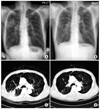
Chest radiography and chest CT during diagnosis and more than 2 months after treatments. (A, C) In a 67-year-old man patchy airspace consolidation in the superior segment of RLL was detected and confirmed lung abscess by chest CT. (B, D) Fifteen months later, the lesion is resolved except for the fibrosteaky density. CT: computed tomography.

Figure 2
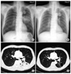
(A, C) In a 64-year-old man with underlying lung cancer in the LUL, segmental air space consoilidation with irregular cavitation is noted in the superior segment of the LLL which is associated with mild adjacent fissural bulging contour. (B, D) Two months later, previous lesion in LLL is improved with a mild residual scar and a pneumatocele. LUL: left upper lobe; LLL: left lower lobe.

Figure 3
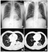
(A) In a 52-year-old man Segmental air space consolidation with peripheral ground glass attenuation is noted in the superior segment of the LLL and lesser extent in the posterior segment of LLL with mild lung volume expansion. (B) Two months later, there is a decrease in the extent of segmental air space consolidation with peripheral ground glass attenuation in previous area. (C) Irregular shaped low density necrotic areas and multiple cavities were noted within the consolidations. (D) But irregular shaped multiple cavities are increased within the consolidations. Newly appeared cicatricial bronchiolectasis and bronchiectasis is seen. LLL: left lower lobe.

References
1. Bartlett JG. Anaerobic bacterial infections of the lung. Chest. 1987. 91:901–909.
2. Hirshberg B, Sklair-Levi M, Nir-Paz R, Ben-Sira L, Krivoruk V, Kramer MR. Factors predicting mortality of patients with lung abscess. Chest. 1999. 115:746–750.
3. Stark DD, Federle MP, Goodman PC, Podrasky AE, Webb WR. Differentiating lung abscess and empyema: radiography and computed tomography. AJR Am J Roentgenol. 1983. 141:163–167.
4. Williford ME, Godwin JD. Computed tomography of lung abscess and empyema. Radiol Clin North Am. 1983. 21:575–583.
5. Wagley PF, Fisher AM, Block AJ. Primary pulmonary abscess. Trans Am Clin Climatol Assoc. 1970. 81:51–56.
6. Snow N, Bergin KT, Horrigan TP. Thoracic CT scanning in critically ill patients. Information obtained frequently alters management. Chest. 1990. 97:1467–1470.
7. Jedrychowski W, Krzyzanowski M. The effect of acute broncho-pulmonary infections on the FEV1 change in 13-year follow-up. The Cracow Study. Eur J Epidemiol. 1990. 6:20–28.
8. Tice AD, Rehm SJ, Dalovisio JR, Bradley JS, Martinelli LP, Graham DR, et al. Practice guidelines for outpatient parenteral antimicrobial therapy. IDSA guidelines. Clin Infect Dis. 2004. 38:1651–1672.
9. Irwin RS, Garrity FL, Erickson AD, Corrao WM, Kaemmerlen JT. Sampling lower respiratory tract secretions in primary lung abscess: a comparison of the accuracy of four methods. Chest. 1981. 79:559–565.
10. Wang JL, Chen KY, Fang CT, Hsueh PR, Yang PC, Chang SC. Changing bacteriology of adult community-acquired lung abscess in Taiwan: Klebsiella pneumoniae versus anaerobes. Clin Infect Dis. 2005. 40:915–922.
11. Wali SO, Shugaeri A, Samman YS, Abdelaziz M. Percutaneous drainage of pyogenic lung abscess. Scand J Infect Dis. 2002. 34:673–679.
12. Bartlett JG. Anaerobic bacterial infections of the lung and pleural space. Clin Infect Dis. 1993. 16:Suppl 4. S248–S255.
13. Levison ME. Anaerobic pleuropulmonary infection. Curr Opin Infect Dis. 2001. 14:187–191.
14. Fifer WR, Husebye K, Chedister C, Miller M. Primary lung abscess. Analysis of therapy and results in 55 cases. Arch Intern Med. 1961. 107:668–680.
15. Rumbaugh IF, Prior JA. Lung abscess: a review of forty-one cases. Ann Intern Med. 1961. 55:223–234.
16. Prasad US, Tiwary A. Scar cancer of the lung. Indian J Chest Dis Allied Sci. 1989. 31:125–128.
17. Auerbach O, Garfinkel L, Parks VR. Scar cancer of the lung: increase over a 21 year period. Cancer. 1979. 43:636–642.
18. Bobba RK, Holly JS, Loy T, Perry MC. Scar carcinoma of the lung: a historical perspective. Clin Lung Cancer. 2011. 12:148–154.
19. Ardies CM. Inflammation as cause for scar cancers of the lung. Integr Cancer Ther. 2003. 2:238–246.


 PDF
PDF ePub
ePub Citation
 Print
Print


 XML Download