

 PDF
PDF ePub
ePub Citation
 Print
Print


Introduction
Blind transbronchial needle aspiration (Blind TBNA) and endoscopic ultrasound guided fine needle aspiration (EUS-FNA) are used to diagnose mediastinal lymph nodes metastases because mediastonoscopy requires general anesthesia. However, the sensitivity and the negative predict value of blind TBNA are known to be 76% and 71% which indicates the low accuracy. Moreover, the sensitivity and the negative predict value of EUS-FNA are known to be 88% and 77%. The accuracy of EUS-FNA seems to be higher but the accessible area for the procedure is limited to the left posterior lymph nodes due to the anatomical location of esophagus1,2. Endoscopic ultrasound-guided fine needle aspiration (EBUS-TBNA) was developed to overcome these limitations of blind TBNA and EUS-FNA in late 2000 and has been used widely since then. This is the real-time lymph node biopsy with 22 G needles through the linear array probe installed in bronchoscopy. This procedure has reduced the complications related to general anesthesia and surgical procedures, and increased accessibility to subcarinal and parahilar lymph nodes where it was difficult to approach with thoracoscopy.
The value of EBUS-TBNA in establishing the stage of lung cancer was proved in previous studies and Nakajima et al. proved that EBUS-TBNA was better to diagnose sarcoidosis than transbronchial lung biopsy (TBLB) and bronchoalveolar lavage (BAL)3. The previous study in a country with low prevalence of tuberculosis reported 46~63% of microbiologic diagnosis rate and greater than 80% of pathological diagnosis rate for intrathoracic tuberculous lymphadenitis when EBUS-TBNA was performed4. Cancer patients are known to possess a higher risk of tuberculosis5-7. Unexpected tuberculous lymphadenitis was found in about 5.1% of lung cancer patients after Solak et al.8 performed mediastinal lymph node biopsy using mediastinoscopy in Turkey and none of the patients were presented with any findings indicating tuberculous lymphadenitis in sputum culture and bronchoscopy. It is necessary to do pathological diagnostic examinations such as mediastinoscopic biopsy because it is 15~25% more probable to misdiagnose the stage of non-small cell lung carcinoma higher than the actual stage, when predicted with positron emission tomography (PET) due to the high lymphatic uptake in a tuberculosis endemic country9,10. Therefore, the possibility of tuberculous lymphadenitis should be considered. Currently, there is not any research conducted regarding the utility of EBUS-TBNA to diagnose tuberculous lymphadenitis in a tuberculosis endemic country, South Korea. Therefore, this study was aimed to examine the value of routine culture for tuberculosis from EBUS-TBNA performed to distinguish tuberculous lymphadenitis from lung cancer or sarcoidosis in the same way of the routine culture for tuberculosis with bronchial washing to increase the diagnosis rate of tuberculosis.
Materials and Methods
1. Subjects
This prospective observation study was conducted with outpatients and inpatients attended (total 86 patients) in Severance Hospital between March 2010 and March 2011 by performing routine culture for tuberculosis from EBUS-TBNA. EBUS-TBNA was performed to differentiate between malignancy and the other benign disease when the causes of the enlarged lymph nodes larger than 1 cm of its axis identified in mediastinum and pulmonary hilum on chest computed tomography (CT) scan were not obvious .The patients who were at high risk for fiberoptic bronchoscopy and those suffered from severe pulmonary disease, cardiac condition, liver disease, neurological defect or bleeding disorder were excluded.
2. Methods
EBUS-TBNA was performed with an endobronchial ultrasound (model BF-UC260F; Olympus, Tokyo, Japan) and a 22-gauge needle (NA-201SX; Olympus) after fiberoptic bronchoscopy (model BF-1T260, BF-260, or FB-6C260, Olympus, Japan). Specimen of all subject collected by bronchial washing were tested for microbiology and cytology examination. The core tissues were obtained from lymph nodes by EBUS-TBNA, were fixed with 10% formalin, were made in slide and were reviewed by pathologists. The rest of the aspirated materials were smeared on the glass slides and fixed with 95% alcohol for cytology examinations. In addition, the aspirated specimens were tested for acid-fast bacillus (AFB) stain and mycobacterium tuberculosis culture using MGIT-BACTEC 960 and ogawa. At the same time, the core tissues were tested for mycobacterium culture if they were aspirated.
Tuberculous lymphadenitis was diagnosed either when granulomatous inflammation accompanying caseous necrosis was confirmed by core tissue examinations and histopathologic examinations or when there were positive results of AFB stain and mycobacterium tuberculosis culture as well as tuberculosis was identified in other areas and the enlarged lymph nodes were improved by tuberculosis treatment. Sarcoidosis was diagnosed when there were correspondent clinical symptoms, the specimen culture for mycobacterium tuberculosis and other bacteria was negative, the causes of allergy, occupational disease and malignancy were excluded and non-caseating epithelioid cell granulomata was pathologically confirmed3. This study was approved by Severance Hospital Institutional Review Board (4-2010-0050).
Results
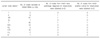
EBUS-TBNA was performed on 86 patients with suspected perihilar and mediastinal lymphadenopathy (total 135 lymph nodes). The mean age of the subjects were 62.4 years old and 63 (73.3%) of them were male and 23 (26.7%) of them were female. Average 1.6 lymph nodes per person were underwent EBUS-TBNA and the locations of lymph nodes were right paratracheal nodes (47, 34.8%), subcarinal lymph nodes (40, 29.6%) and right hilar lymph nodes (14, 10.4%) in the order of frequency (Table 1).
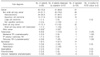
The final diagnoses of the subjects were cancer in 62 patients (44 non-small cell carcinomas, 12 small cell carcinomas, 6 other types of tumors), 1 lung cancer accompanying tuberculosis, 7 cases of tuberculosis, 7 cases of sarcoidosis, 2 cases of aspergillosis and 2 cases of pneumoconiosis. Among total 63 cancer patients, the sensitivity of EBUS-TBNA aspirated specimen cytology test was 80.9% and the sensitivity of core tissue pathology test was 76.2% (Tables 2, 3). Two of 7 sarcoidosis patients were at the stage I without any lung parenchymal lesions and the rest of 5 patients were accompanied with lung parenchymal lesions. One of them was diagnosed by TBLB and EBUS-TBNA, another patient was diagnosed by mediastinoscopy and the rest of 5 patients were diagnosed by EBUS-TBNA. The pathological findings corresponded to sarcoidosis were found in 8 aspirated specimens (57%) and 7 core tissues (70%).
There were 8 confirmed tuberculosis patients and one of them was accompanied with lung cancer. One of them (12.5%) was diagnosed by EBUS-TBNA aspirated specimen cytology test and 3 of them were diagnosed after the observation of caseous necrosis on pathologic examinations of core tissues (37.5%). Among the patients with the positive mycobacterial culture result, 2 of them were confirmed in aspirated specimens (25%) and 2 of them were confirmed in core tissues (25%) (Tables 2, 3). Polymerase chain reaction test for M. tuberculosis (TB-PCR) was also performed to total 8 cases of confirmed enlarged lymph nodes secondary to tuberculosis and one of them presented with a positive result showing 12.5% of sensitivity. Performing TB-PCR to 75 patients with the other diagnosis apart from tuberculosis, all the patients were presented with a negative result indicating 100% specificity. Among 4 patients presented with correspondent findings to tuberculosis on EBUS-TBNA, 2 of them were mediastinal lymphadenopathy without lung parenchymal lesions and the other 2 patients accompanied with lung parenchymal lesions additionally had mediastinoscopy and TBLB to aid the diagnosis. In detail, a case of tuberculosis was confirmed by core tissue histopathology test but mycobacterial culture was negative and another patient showed positive TB-PCR and presented with caseating granuloma on histopathologic examinations of TBLB of core tissues and lung parenchyma. The diagnosis of another patient was confirmed by mycobacterial culture of lymph node tissue, aspirated specimen and core tissue. There was only one patient who was diagnosed with tuberculosis by only mycobacterial culture without the pathologic confirmation of EBUS-TBNA aspirated specimen examinations. The patient was already confirmed as tuberculosis by pathologic examination through mediastinoscopy before it was confirmed by the mycobacterial culture result. Among 4 patients who were diagnosed with tuberculosis despite of the negative EBUS-TBNA result, one of them was diagnosed by thoracentesis and pleural biopsy. The other 3 patients were clinically diagnosed with tuberculosis through imaging tests, clinical symptoms, and improvement after administrating anti-tuberculosis medications. The tuberculosis accompanied with lung cancer was confirmed by AFB smear and started tuberculosis therapy prior to the admission but there was no evidence of tuberculosis on EBUS-TBNA (Figure 1). There were not any complications related to EBUS-TBNA such as bleeding or pneumothorax.
Discussion
About 50% of extrapulmonary tuberculosis is tuberculous lymphadenitis and it is the most common in childhood tuberculosis, but it has been increasing among adults in recent years. Tuberculous lymphadenitis is the most prevalent in tuberculosis endemic country and it has been reported that it is most commonly seen in female and in the third to fifth decade domestically11. Although fine needle aspiration is usually performed to confirm tuberculous lymphadenitis12, the results can be ambiguous in many cases and its sensitivity is usually low. Moreover, specific characteristics are not identified in immunosuppressed patients such as acquired immune deficiency syndrome even if granuloma is observed. Therefore, excisional biopsy is necessary in some cases.
Intrathoracic tuberculous lymphadenitis is shown in 5~7% of adult pulmonary tuberculosis and 2% of them are diagnosed without lung parenchyma lesions resulting low diagnostic rate with bronchoscopy13. Chest CT finding of tuberculous lymphadenitis is radiolucency in the middle of lymph nodes created by necrosis and enhancement in surrounding area indicating histopathological caseous necrosis. However, these findings are observed in only in 33~75% of patients, so chest CT cannot be used for a definite diagnostic method14.
Malignancy is usually suspected when the maximum standard uptake value (SUV) on PET is greater than 2.5 but the value increases to the maximum level when there is active tuberculosis. Therefore, active tuberculosis cannot be differentiated from malignancy only by SUV and its specificity is low despite its high sensitivity15,16. For this reason, the pathological diagnosis was necessary to diagnose tuberculous lymphadenitis and the diagnostic value of fine needle aspiration was reported in a number of researches. According to the study of Ellison et al.17 conducted in the United States with less prevalence of tuberculosis, there were 58 AFB smear positives (24.4%) and 82 mycobacterium culture positives (34.4%) performing needle aspiration from 238 lymph nodes of 180 tuberculosis patients and the sensitivity was increased to 53% when AFB smear, mycobacterium culture and pathologic findings of granuloma were combined. Nayak et al.18 conducted a research performing fine needle aspiration to 21 human immunodeficiency virus (HIV) positive patients and 21 HIV negative patients accompanied with tuberculous lymphadenitis and reported that the positive AFB smear, the positive mycobacterium culture and the positive pathological findings correspondent to tuberculosis were 61.9%, 23.8% and 14.3%, respectively in HIV positive patients, and 9.5%, 38.1% and 52.4% in HIV negative patients. These results suggested that the need for additional histological tests was higher in HIV negative patients than HIV positive patients.
Ayed and Behbehani13 performed bronchoscopy and mediastinoscopy to isolated thoracic tuberculous lymphadenitis patients without lung parenchymal lesions at the same time. They reported that all tuberculous lymphadenitis was identified by mediastinoscopy but none of the mycobacterium culture from sputum identified tuberculosis and only 9% of the mycobacterium culture from bronchial washing identified tuberculosis. Chang et al.19 reported that tuberculosis was diagnosed from 36% of sputum culture, 90% of peripheral lymph node biopsy and 75% of bronchoscopy among the pulmonary tuberculosis patients accompanied with tuberculous lymphadenitis. Cetinkaya et al. diagnosed 8 tuberculosis patients among 10 tuberculosis patients (80%) with only fine needle aspiration without any other diagnostic investigations20.
Cancer patients carry 9 times higher risk of tuberculosis than general population4 and the prevalence of tuberculosis among lung cancer patients is reported to be 916 per 100,000 people (MSK series)5 or 901 per 100,000 people (Gopalakrishnan)6. About 5.1% of lung cancer patients accompany with tuberculous lymphadenitis7 and 82.4% of bronchogenic carcinoma patients accompanied with tuberculosis also show mediastinal lymphadenopathy21. Impaired cell-mediated immunity due to the characteristics of tumors themselves or chemotherapy reactivates pulmonary tuberculosis and the survival rate of lung cancer patients with active tuberculosis is lower than those without22. Simultaneous tuberculosis therapy with chemotherapy is important in lung cancer patients because it is known that tuberculosis does not affect the clinical progress of lung cancer when it is treated appropriately23.
EBUS-TBNA has been developed to establish the mediastinal stage and carries less risk of neurovascular damage and infection than surgical methods under general anesthesia24. Kuo et al. performed EBUS-TBANA on lung cancer patients accompanied with enlarged lymph nodes less than 1cm in the tuberculosis endemic area and reported 80.6% sensitivity of EBUS-TBNA, 100% specificity, 100% positive predictive value and 85.7% negative predictive value to diagnose the tumor lymphatic invasion. This results showed the more superior results of EBUS-TBNA than 18% specificity and 44% positive predictive value of PET-CT25. Marcus et al. performed EBUS-TBNA to 153 patients suspected of mediastinal malignancy and confirmed sarcoidosis in 5.2% (8/153) of them26 and Nakajima proved that the diagnostic accuracy of EBUS-TBNA was higher than that of BAL or transbronchial lung biopsy for the patients suspected of sarcoidosis3. Therefore, EBUS-TBNA became a standard diagnostic method for malignancy and sarcoidosis but the researches regarding its diagnostic value for tuberculous lymphadenitis only started to be conducted in recent years4,27. EBUS-TBNA is a useful method for the patients suspected of tuberculous lymphadenitis located in inaccessible place for the biopsy because it facilitates the diagnosis and drug sensitivity of cultured tuberculosis can be tested through aspirated specimen and pathologic specimen of core tissue.
Among 8 patients with negative results of AFB smear and mycobacterium culture of sputum and bronchial washing, four patients were diagnosed either histologically or by mycobacterium culture from EBUS-TBNA This result was correspondent to the conclusions in previous studies stating that EBUS-TBNA was useful to diagnose tuberculous lymphadenitis although the sensitivity of mycobacterium culture and pathology tests of EBUS-TBNA was low in this study. However, the additional mycobacterium culture of aspirated specimen was not beneficial because only one case (1.4%) was diagnosed by mycobacterium culture of aspirated specimen and core tissue from lymph node and tuberculous lymphadenitis was already diagnosed by mediastinoscopy before the culture result was available.
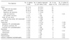
Only 1.5% of cancer patients accompanied with tuberculosis in this study whereas 5.2% of cancer patients accompanied with tuberculosis in the study of Solak. The reasons for lower incidence are small sample size and that additional surgical mediastinal lymph node biopsy was not performed. Moreover, the selection bias should have been occured during lymph nodes selection for EBUS-TBNA. The utility of EBUS TBNA was underestimated as a diagnostic tool for tuberculous lymphadenitis because EBUS TBNA was usually used for lymph node biopsy in cancer patients such as lung cancer in tertiary hospital. The sensitivity and the specificity of mycobacterium culture and pathology tests from EBUS-TBNA were 50% and 100% respectively which were lower than 95% and 100% in the study of Hassan et al.27. The sensitivity seems to be low because this study was conducted with mediastinal lymphadenitis including cancer patients and a number of inactive lymph node lesions whereas the previous studies included the patients who were strongly suspected of intrathoracic tuberculous lymphadenitis without lung parenchyma lesions. The higher mycobacterium culture was expected if the subjects were limited to the patients with enlarged mediastinal lymph nodes or to the patients suspected of tuberculous lymphadenitis presented with radiolucency in the middle of lymph nodes. The sensitivity of EBUS-TBNA was 100% as the diagnosis of all 4 isolated mediastinal lymphadenopathy in this study. This result is similar to bigger scale of study on intrathoracic tuberculous lymphadenitis patients without lung parenchyma lesions presented with negative AFB smear and mycobacterium culture reporting 94% of sensitivity4. However, the results of the previous study is not feasible to apply in South Korea with high prevalence of tuberculosis and low prevalence of HIV because they were conducted in the countries where the prevalence of tuberculosis was low and HIV positive patients were included. It is thought to be beneficial to conduct large scale of researches in tuberculosis endemic countries in order to examine the clinical role of EBUS-TBNA for tuberculosis lymphadenopathy.
In conclusion, EBUS-TBNA is beneficial for isolated mediastinal lymphadenopathy which needs to exclude tuberculous lymphadenopathy when sputum collection is difficult or AFB smear is negative, but the advantage of additional routine AFB smear and mycobacterium culture of fine needle aspiration specimen to diagnose tuberculous lymphadenitis accompanied with malignancy is not justified.
 XML Download