

 PDF
PDF ePub
ePub Citation
 Print
Print


Introduction
The lung is the most frequent site of breast, renal, colorectal, or melanoma metastasis. However, isolated pleural metastasis without lung parenchymal metastasis also can occur in some cases. In such cases, specimens are usually obtained from pleural fluid aspiration cytology, percutaneous needle biopsy, or surgical resection of the pleura1,2. This case study describes metastatic renal cell carcinoma of the pleura that was diagnosed by endobronchial ultrasound-guided transbronchial needle aspiration (EBUS-TBNA). To our knowledge, this is the first case of EBUS-TBNA for a pleural mass.
Case Report
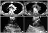
A 51-year-old non-smoking male with no chronic disease presented with a cough and right-side chest pain for 1 month. He visited another institution, where a chest x-ray showed a widened mediastinum and pleural effusion in the right hemithorax. Contrast-enhanced computed tomography (CT) of the chest revealed a small pleural effusion and thickened pleura forming multiple masses (Figure 1A). There was no parenchymal lesion in either lung. On positron emission tomography (PET)/CT, 18F-fluorodeoxyglucose (FDG) uptake was noted in the pleural masses, which were suspected to be malignant mesothelioma or metastatic tumor (Figure 1B). A left renal mass with FDG uptake was suspicious of renal cell carcinoma (RCC). The patient underwent a percutaneous needle biopsy of the pleura. A few atypical mesothelial cells were seen in the tissue specimen, which was insufficient for immunohistostaining. The percutaneous pleural biopsy was repeated, but again, the specimen was insufficient for a diagnosis. Therefore, the patient was referred to Samsung Medical Center (Seoul, South Korea) for a histopathological diagnosis.
Because one pleural mass was adjacent to the right main bronchus, we decided to perform EBUS-TBNA of the pleural mass using a flexible convex probe ultrasonic puncture bronchoscope with a 7.5-MHz-frequency linear scanning transducer (CP-EBUS, BF-UC206F-OL8; Olympus, Tokyo, Japan). A dedicated ultrasound scanner (EU-C2000; Olympus Tokyo, Japan) was used as the image processor. On flexible bronchoscopic examination, the posterior wall of the right main bronchus was narrowed by extrinsic compression. On EBUS, a huge, heterogeneous, low-echogenic pleural mass was seen at the right main bronchus level (Figure 1C). We performed EBUS-TBNA of the pleural mass and obtained a large tissues core (Figure 1D). The patient did not suffer any complication from the procedure such as pneumothorax, pulmonary infection, or hemoptysis.
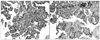
The specimens from the pleural mass were sufficient for histopathological examination and showed papillae formation of cells with atypical nuclei (Figure 2A).
Immunohistochemical staining (IHC) was negative for calretinin, Wilm's tumor-1 (WT-1), and Hector Battifora mesothelial cell-1 (HBME-1), which are markers for malignant mesothelioma. It was also negative for IHC staining for transcription termination factor-1 (TTF-1), indicating a low probability of peripheral adenocarcinoma of the lung. The specimen was positive for CD10 (Figure 2B). In this case, the patient was suspected of having RCC on PET/CT, and the positive reaction for CD10 suggested metastatic malignancy from RCC3,4. Four days after the EBUS-TBNA, a renal biopsy was performed, and the pathology indicated papillary-type RCC. Therefore, the patient was diagnosed with papillary RCC with metastasis to the right pleura.
Discussion
Pleural metastasis without lung parenchymal spread from extrathoracic malignancy is rare and difficult to diagnose. As Mitsuhiro et al. reported, it can occur several years after curative resection, especially in patients with RCC. It is often confused with malignant mesothelioma because the CT images are similar5,6. To distinguish metastatic malignancy from malignant mesothelioma, it is important for clinicians to obtain a sufficient specimen for histopathological examination.
Endobronchial ultrasound-guided transbronchial needle aspiration is a very useful procedure for obtaining specimens from mediastinal and hilar lymph nodes or lung masses accessible from airways in both primary lung cancer and metastatic malignancy7. It has high diagnostic accuracy and excellent safety profiles, with minimal invasiveness under real-time visualization8. EBUS-TBNA can also be performed in other specific cases. Nakajima et al.9 reported a case of central airway stenosis caused by a mediastinal cyst, which improved after cyst aspiration using EBUS-TBNA. Takahiro et al. also reported a diagnosis of spinal chondrosarcoma based on EBUS-TBNA10; as the protruding posterior mediastinal mass was adjacent to the carina, a histopathological diagnosis could be made using EBUS-TBNA. These cases show the usefulness of EBUS-TBNA for the diagnosis of mediastinal masses and the therapeutic utility of EBUS-TBNA for the aspiration of mediastinal cysts. Nevertheless, there has been no report of EBUS-TBNA for the diagnosis of pleural disease.
This case study reports a renal cell carcinoma with pleural metastasis. What is more unusual about our case is that a specimen was obtained from the pleural mass using EBUS-TBNA. To our knowledge, this is the first case of EBUS-TBNA for a pleural mass. Although the patient underwent percutaneous needle biopsy twice, the specimens were insufficient for IHC staining, which is necessary for a histopathological diagnosis. We decided to perform another diagnostic modality rather than a third percutaneous needle biopsy. EBUS-TBNA, instead of a surgical biopsy such as video-assisted thoracoscopic surgery, ensured that the patient was free from pain and did not require general anesthesia, and the procedure was less invasive. Furthermore, sufficient tissues from the EBUS-TBNA enabled the IHC examination of multiple markers, which helped make a definite histopathological diagnosis in this patient.
Posterior pleural masses or lesions which are located in the proximity to both main bronchi or right bronchus intermedius can be accessible by EBUS-TBNA. Respiratory physicians should aware that it is possible to obtain an adequate specimen from a pleural mass safely using EBUS-TBNA when the mass is adjacent to an airway.
 XML Download