

 PDF
PDF ePub
ePub Citation
 Print
Print


Introduction
Pneumocystis species were initially regarded as protozoa and are now classified as fungi based on DNA sequence analysis of srRNA genes1. The Pneumocystis species that infects humans was recently renamed Pneumocystis jirovecii, while the old name, P. carinii, is reserved for species that infect other animals. P. jirovecii is a pathogen that has become an increasingly important cause of opportunistic infection2. So far, definite diagnosis of P. jirovecii pneumonia (PJP) requires morphological identification of the organism because serologic tests are not reliable and culture of the pathogen is very difficult3. Because most patients with PJP are immunocompromised, diagnosis without invasive procedures is favored in order to avoid unnecessary complications. Although transbronchial lung biopsy (TBLB) has an equivalent yield for the detection of PJP as bronchoalveolar lavage (BAL) cytology, it is associated with a greater frequency of complications, such as hemorrhage and pneumothorax, especially in patients with human immunodeficiency virus (HIV)3. Therefore, noninvasive methods with high sensitivity for the diagnosis of PJP, such as BAL fluid cytology, are required for better management of PJP patients.
In this study, we reviewed BAL fluid cytology from PJP cases seen at our institution between 2004 and 2010 that were proven to be PJP by surgical biopsy. In this report, we summarize the clinical status and findings of BAL fluid cytology of these patients and evaluate the sensitivity of BAL fluid analysis for diagnosis of PJP.
Materials and Methods
1. Patients
This study included 30 patients with PJP who were diagnosed through surgical biopsy, including TBLB (29 cases) and video-associated thoracoscopic surgery (VATS) assisted lung biopsy (one case). Clinical data were collected by retrospective review. Twenty-five patients with pulmonary infiltrates on radiology without evidence of PJP were enrolled as the control group. The control group was consisted of 16 with community acquired pneumonia, 7 hospital acquired pneumonia and 2 with acute respiratory distress syndrome associated with malignancy and hemodynamic disorder.
2. Review of BAL fluid
The BAL fluids analyzed in this study were prepared with the conventional cytospin method. Hematoxylin and eosin (H&E) stain was used for all specimens. Gomori's methenamine silver (GMS) staining and immunohistochemical staining for P. jirovecii were performed in only three and one case(s), respectively.
Two pathologists (JY Sung and J Han) reviewed the BAL fluid samples from both the PJP and control cases. The concordance rate between the initial cytologic diagnosis and review diagnosis was also evaluated. All PJP and control cases were reviewed for cytologic findings, including low power pattern, frothy exudates, hemosiderin- laden macrophages, fibrinoid materials, and the kinds of infiltrated inflammatory cells. For the parameters of hemosiderin-laden macrophages, fibrinoid materials, and the kinds of infiltrated inflammatory cells, we classified "easily found" (with the proportion of more than 25% of all of the cell population in BAL fluid) and "rare" (less than 25%).
3. Statistical analysis
We used Statistical Product and Services Solutions (SPSS version 18.0; SPSS Inc., Chicago, IL, USA) statistical software to carry out the statistical analysis. The Pearson χ2 test and Fisher's exact test (for analysis of frothy exudates) were used to identify the cytologic findings that showed statistically significant differences between the PJP and control groups. The tests were two-sided. Differences were considered statistically significant when the p-value was <0.05.
Results
1. Patient characteristics
The patient characteristics of the 30 PJP cases are summarized in Table 1. The mean age was 48.8±2.3 years (range, 24.0~72.0; data shown as median±standard error) and 10 of the 30 patients were female. All patients with PJP were immunocompromised. The underlying clinical conditions of PJP patients were as follows: HIV positive, 13 (43.3%); hematologic malignancy and chemotherapy, 6 (20.0%); organ transplantation, 9 (30.0%); and autoimmune disease and steroid therapy, 2 (6.6%). Seven of the 30 PJP patients were also diagnosed with diffuse alveolar damage (DAD) in addition to PJP.
2. Result of review of cytologic findings in PJP patients
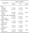
Table 2 summarizes comparisons of the cytological findings between the PJP patients and the control group. In the BAL fluid samples from PJP patients, frothy exudates were identified (p<0.001) whereas none of the control cases showed frothy exudates. And rare neutrophils were present (p=0.023) in those from PJP patients. The rest of the cytological findings, such as low power pattern, easily detected hemosiderin- laden macrophages, fibrinoid materials, and lymphocytes, were not correlated with PJP.
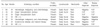
Among 30 patients with PJP, seven patients were diagnosed with DAD with associated PJP (Table 3). The DAD patients had different findings in their BAL fluid. Two (28.6%) showed no frothy exudates, which are the characteristic finding of PJP. Therefore, we were unable to make diagnoses of PJP from the BAL fluid of these two patients.
Discussion
A study from the United States reported that PJP was the most prevalent opportunistic disease4. According to Choe et al.5, who analyzed opportunistic infections in HIV-infected patients in Korea between 1985 and 1998, tuberculosis was the most frequent opportunistic infection (25%), followed by candidiasis (21%), herpes zoster (20%), and PJP (10%). Cho et al.6 reported that the most prevalent (21.6%) opportunistic disease in HIV-infected patients from 1985 to 2000 was candidiasis, followed by PJP (15.9%), tuberculosis (12.5%), and cytomegalovirus (CMV) infection (9.1%).
Although Pneumocystis is one of the important causes of opportunistic infection, there are presently few reviews of data regarding PJP in Korea. In our study, the patients with PJP had immunodeficiency associated with AIDS (13, 43.3%), organ transplantation (9, 30.0%), hematologic malignancy (6, 20.0%), and autoimmune disease (2, 6.6%). AIDS was the most frequent predisposing factor, but organ transplantation and malignancy are also common risk factors for PJP.
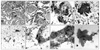
As shown in Figure 1, the characteristic findings of PJP for both cytologic and surgical specimens are foamy alveolar casts called frothy exudates3,7,8. When this finding was noted, we can diagnose the case as PJP. The term of "honeycomb exudates" also used to describe this finding and the honeycomb texture is derived from the presence of the PJP cysts in the intra-alveolar proteinaceous material. With statistical significance, the frothy exudate was found in 93.3% of PJP whereas none of control case showed it. So, the diagnostic value of frothy exudates for PJP is reconfirmed in our review of BAL fluid. Additionally, we found that BAL fluid from PJP patients has a tendency to have rare neutrophils in comparison with a group of pneumonia patients without evidence of PJP.
BAL fluid cytology is easy to prepare and can yield more rapid results than surgical procedures. Furthermore, the sensitivity for detection of PJP by BAL is known to be high, ranging from 79% to 98%9-11. When we reviewed the BAL fluids of PJP patients in our institute, the sensitivity was 93.3%. However, the sensitivity of the initial interpretation of BAL fluid was 50.0%, and the concordance rate was 56.7%. Because pathologists usually concentrate on ruling out malignancy, an event like this can occasionally occur. Another reason may be a lack of ancillary tests, including special and immunohistochemical stains. Among 30 cases, 27 (14 diagnosed as negative at initial diagnosis and 13 diagnosed as positive at initial diagnosis) did not undergo any special or immunohistochemical staining. Chechani et al.12 previously reported that the diagnostic yield of BAL fluid cytology may be increased by use of special stains, such as Gram-Weigert and GMS stains. Recently, the use of immunohistochemical staining for P. jirovecii is increasing, and it is reported to be helpful in the diagnosis of PJP13. However, when we reviewed our 30 cases, diagnosis of PJP was not difficult even though we did not perform additional special or immunohistochemical staining. Therefore, if pathologists are aware of the possibility of PJP, they can reduce the false negative rate even if special or immunohistochemical staining is unavailable.
Another common histologic manifestation of PJP is DAD, which is most often seen in non-AIDS patients14. In cases of PJP with DAD, intra-alveolar frothy exudates may be focal and scanty and, thus, difficult to find15. The results in our study concur with a previous report. Among 30 cases of PJP in our institute, seven cases presented with DAD and two of these were patients with HIV (Table 3). Two out of seven cases with DAD showed a scanty amount of frothy exudates in surgical specimens. As expected, we were unable to find any diagnostic findings, such as frothy exudates, in BAL fluid from these patients. This result suggests that even if there is no evidence of PJP in the BAL fluid, PJP cannot be excluded in cases of immunocompromised patients with DAD, especially non-HIV infected patients. Clinicians need to be alert to this possibility because the proportion of non-HIV infected immunocompromised patients is increasing.
In this study, we reviewed BAL fluid samples from 30 PJP patients at an institute in Korea. In addition to HIV infection, organ transplantation and malignancy are also important causes of PJP. Especially, in the cases with information of immunodeficiency, we need to be aware of the possibility of PJP and careful cytologic examination should be done. So the communication between pathologist and clinician is very important. For the evaluation for PJP, BAL fluid cytology is recommemded, because it is very rapid than surgical process and has high sensitivity. Finally, in the case of PJP with DAD, we cannot exclude PJP even if there are no frothy exudates in the BAL fluid.
 XML Download