

 PDF
PDF ePub
ePub Citation
 Print
Print


Introduction
Gefitinib and erlotinib, which are epidermal growth factor receptor (EGFR) inhibitors, are useful for treating advanced non-small cell lung cancer patients. However, patients who can benefit from these EGFR inhibitors are limited to about 30% of the total non-small cell lung cancer cases having mutation of EGFR gene1. Moreover, even the lung cancers responsive to the agents eventually develop resistance due to the appearance of secondary EGFR mutations or an activated c-Met signal transduction pathway which is a bypass2.
As for radiation therapy, combination with chemotherapy is positioned as a standard treatment for locally advanced non-small cell lung cancer in stage IIIB. Despite, the current combination therapy still has unsatisfactory treatment results, which often lead to systemic chemotherapy to treat recurrence3.
There have been trials to improve treatment effects by combining these EGFR inhibitors with radiation therapy. One of the examples is the multinational randomized study on the combination of irradiation with cetuximab administration for locally advanced squamous cell carcinoma of the head and neck, which contributed to extended survival periods4. In several animal experiments, EGFR inhibitors were reported to enhance sensitivity of lung cancer cells on radiation5-8, but no independent report on enhanced treatment results from combination therapy of irradiation and administration of EGFR inhibitors has been released yet in lung cancer patients.
According to numbers of studies, EGFR pathways are activated when cancer cells are irradiated, and RAS/mitogen-activated protein kinase (MAPK) pathway and phosphatidylinositol 3-kinase (PI3K)/AKT pathway are also activated and accordingly, cell proliferation occurs to resist against radiation9-12. If EGFR pathways are more activated than the ground state upon irradiation, EGFR inhibitors may be administered at this point of time with an expectation of enhanced effects. Therefore, we developed a hypothesis; if EGFR inhibitors are administered when EGFR pathways are activated upon irradiation, the effects of the agents will enhance. We tried to verify the hypothesis using lung cancer cells.
Materials and Methods
1. Cell culture and reagents
Lung cancer cell strains of A549 and NCI-H460 were purchased from American Type Culture Collection (ATCC; Rockville, MD, USA). The PC-9 cell strain having exon 19 deletion of the EGFR gene was provided by F. Koizumi and K. Nishio (National Cancer Center Hospital, Tokyo, Japan). Each cell strain was cultured using the RPMI1640 culture media containing 10% fetal bovine serum and 1% gentamicin sulfate in a CO2 cell incubator (37℃, 5% CO2). Gefitinib, an EGFR inhibitor, was provided by AstraZeneca Korea (Seoul, Korea). Methylthiazol-2-yl-2, 5-diphenyl-tetrazolium bromide (MTT) and propidium iodide (PI) were purchased from Sigma (St. Louis, MO, USA), and annexin V-FITC was purchased from BD Bioscience (San Jose, CA, USA). Protein assay kit for protein quantification was purchased from Bio-Rad (Richmond, CA, USA). Antibody against caspase-3, and secondary antibody were purchased from Cell Signaling (Boston, MA, USA), and antibody against p-EGFR, p-Akt, p-ERK, PARP and β-actin was purchased from Santa Cruz Biotechnology (Santa Cruz, CA, USA). Enhanced chemiluminescence (ECL) kit was purchased from PerkinElmer (Waltham, MA, USA).
2. Radiation-resistant cell lines
A 2 Gy of gamma ray was radiated to each of A549 and PC-9 cells using Cs-137 cell radiator (Atomic Energy of Canada Ltd, Canada). For establishing resistance, A549 and PC-9 cells had survived from the 2 Gy of gamma irradiation were cultured up to 80% confluence. Cells were repeatedly irradiated with escalating dose of gamma ray and finally, cells were exposed with 6 Gy for 6 months or more to be established as resistant cell lines.
3. Methylthiazol-2-yl-2, 5-diphenyl-tetrazolium bromide (MTT) analysis
6×10³ of cells were seeded in a 96-well plate and cultured for 12 hours or more. And then, gefitinib was treated by each concentration for 72 hours. Three hours after MTT reagent was applied to each plate, 10% of sodium dodecyl sulfate solution was added to dissolve violet formazan generated by living cells. After 24-hour incubation, results were analyzed at 595 nm using microplate reader (Bio-Rad; Richmond, CA, USA).
4. Apoptosis assay
A total of 4×10⁵ cells were cultured on a 60 mm dish one day before gefitinib treatment, and cells were collected after 48 hours. Each cell was treated with annexin V binding buffer (150 mM NaCl, 18 mM CaCl2, 10 nM HEPES, 5 mM KCl, 1 mM MgCl2) and then, reacted with annexin V (1 g/mL) and 50 g/mL propidium iodide (PI) at a dark place for 30 minutes. After then, fluorescence-activated cell sorting (FACS) was conducted and analyzed using CellQuest software (BD Biosciences; Franklin Lakes, NJ, USA).
5. Western blot
In order to confirm changes in cell death and EGFR-related signaling molecules, cultured cells were collected and dissolved in lysis buffer (50 mM HEPES, 150 mM NaCl, 10% glycerol, 1% Triton X-100, 1.5 mM MgCl2, 1 mM EGTA, 1 mM sodium vanadate, 10 mM sodium pyrophosphate, 10 mM NaF, 300µM p-nitrophenyl phosphate, 1µg/mL leupeptin, 1 mM PMSF, 10 µg/mL aprotinin, pH 7.3) and then, centrifuged and quantified. Using same amount of protein, western blot was done with 10% or 12% SDS-PAGE gel. After electrophoresis, gel was moved to nitrocellulose membrane, and soaked in TBST (20 mM Tris-HCl, pH 7.6, 137 mM NaCl, 0.01% Tween-20) solution containing 5% nonfat skim milk for an hour at room temperature. Each of the primary antibodies was diluted to the ratio of 1:1,000 and reacted over night. Membranes which completed reaction were washed 3 times with TBST and then, reacted with the secondary antibody for about an hour and washed 3 times with TBST. The washed membranes were developed using the ECL kit.
Results
1. Comparison of responses of radiation-resistant cells and their parental cells to gefitinib
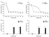
To begin with, we authors used established radiation-resistant cell lines which have been kept for experiments. Gefitinib was administered by each concentration to radiation-resistant cell lines which were repeatedly exposed to radiation and to their parental cells and then, survival rates were calculated through MTT analysis 72 hours later. In case of PC-9 cells having exon 19 deletion of EGFR gene, no significant difference of survival was observed between parental cells and radiation-resistant cells upon administration of gefitinib. By contrast, radiation-resistant A549 cells, not having mutant EGFR genes, showed significant reduction in survival rates compared with parental cells upon administration of gefitinib. Half maximal inhibitory concentration (IC50) of A549 parental cells were 6.10µM, and IC50 of radiation-resistant cells was 0.008µM showing a significant reduction (Figure 1A).
Additionally, 48 hours after gefitinib administration, FACS was conducted using annexin V and PI and apoptosis was analyzed. In case of PC-9 cells, apoptosis fractions induced after administering different concentrations of gefitinib showed no difference between the parental cells and the radiation-resistant cells. By contrast, in case of A549 cells, upon administering 0.1µM of gefitinib, parental cells and radiation-resistant cells showed 4.64% and 45.06% of apoptosis fractions respectively, and with 1µM of gefitinib, 6.52% and 43.13% respectively, showing significant difference likewise cell survival rates (p<0.05) (Figure 1B).
2. Changes in responses to gefitinib after ionizing radiation
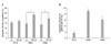
Each of PC-9, A549, NCI-H460 cells was irradiated with 2 Gy and after 48 hours, 1µM, 20µM and 30µM of gefitinib, being equivalent to IC10~IC20, were administered. FACS analysis was conducted 48 hours after the administrations to measure apoptosis fraction.
In case of PC-9 cells, gefitinib-induced apoptosis fractions were 15.19% without previous radiation, and 16.81% with previous radiation, showing no significant difference between the treatments. By comparison, A549 cells showed 14.16% and 23.06% (p<0.05) with the respective treatment, and NCI-H460 cells showed 13.12% and 19.16% (p<0.05) with the respective treatment, showing significant differences (Figure 2A). The difference of gefitinib-induced apoptosis fractions between the treatment with previous radiation and the treatment without previous radiation showed 1.62% with PC-9 cells, 8.90% with A549 cells, and 6.04% with NCI-H460 cells (p<0.05) (Figure 2B).
3. Changes in apoptosis-related proteins
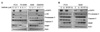
In order to confirm whether the changes observed in the above experiments were also found in apoptosis-related proteins or not, western blot was conducted 24 hours after administration of gefitinib. According to the experiments using radiation-resistant cells and their parental cells, no significant difference in reduction of procaspase 3, increase in caspase 3 active form and PARP cleavage, which are expected to occur upon administration of gefitinib, was found between radiation-resistant and parental cells in case of PC-9 cells. By contrast, in case of A549 cells, radiation-resistant cells showed comparatively more changes of decrease in procaspase 3, increase in caspase 3 active form and PARP cleavage than parent cells did upon administration of gefitinib (Figure 3A).
In the experiment of gefitinib treatment after radiation, PC-9 cells showed no significant difference between the treatment without previous radiation and the treatment with previous radiation, but A549 cells showed significant changes of decrease in procaspase 3, increase in caspase 3 active form and PARP cleavage in case of the gefitinib treatment with previous radiation compared with that without previous radiation (Figure 3B).
Discussion
Based on the above results, EGFR wild-type A549 and NCI-H460 cells showed a significantly enhanced effect of gefitinib after irradiation. However, PC-9, having EGFR mutation, did not show these results. This is interesting results which have not been expected. It has been well known that wild-type cells without EGFR mutation generally show a resistance against EGFR inhibitors. If effects of EGFR inhibitors significantly increase after irradiation, we could expect effects of EGFR inhibitors even in these EGFR wild type cells.
A number of studies reported activated EGFR pathways upon irradiation on cancer cells resulting in cell proliferation and resistance against radiation effects. Mechanisms involved in the above are firstly, swift repopulation through activated RAS/MAPK/ERK pathways after the irradiation9,13-16 and secondly, increased cell survival due to activated PI3K/AKT pathways17,18. Thirdly, a hypothesis explains that EGFR directly involves in repairing radiation-caused DNA damage as a mediator. According to studies, when A549 cells were irradiated, EGFR moves into nucleus to combine with protein kinase (DNA-PK) which is essential for repairing DNA damaged by radiation10.
In other words, this implies that activated EGFR pathways upon irradiation possibly functions as a mechanism of cancer cell survival. The fact that the EGFR pathway is activated compared with a ground state after irradiation may become a base to setup a hypothesis explaining increased responses to EGFR inhibitors. However, further studies are necessary to explain the reason why no same result is shown in PC-9 cells with EGFR mutation. As one of the possible explanations, PC-9 cells with EGFR mutation already have sufficiently activated EGFR pathways and accordingly, additional activation stimulation by irradiation may not result in much difference.
In the present study, radiation-resistant cell lines which were already exposed to radiation were compared with their parental cells. Meanwhile, to find out effects of gefitinib, the agent was administered to each of the irradiated and non-irradiated cells. In both experiments, they showed the same results, even though the degree of irradiation was much different from each other experiment.
Most of the studies on EGFR inhibitors and radiation therapy are focused on finding out a radiosensitizer which can improve the treatment effect of radiation therapy by pre-processing EGFR inhibitors5-8,19-24. There is an example of multinational randomized study reporting a statistically significant increase in survival periods by combining cetuximab with radiation therapy for treating locally advanced squamous cell carcinoma of head and neck. According to the above study, a group of single treatment with 213 participants and a group of combined treatment with 211 participants showed a statistically significant difference with 14.9 months and 24.4 months respectively in terms of the duration of locoregional control (p=0.005). In addition, the overall survival periods were 29.3 months and 49.0 months respectively, and the progression-free survival periods were 12.4 months and 17.1 months respectively, which were also statistically significant4. Being different from the results of the above study, the present study showed that previous radiation can change the effects of EGFR inhibitors which were administered following irradiation. In conclusion, the significance of the present study is suggesting that irradiation may increase the effects of EGFR inhibitors.
 XML Download